. 1998 Sep 21;188(6):1203-8.
doi: 10.1084/jem.188.6.1203.

High frequency of skin-homing melanocyte-specific cytotoxic T lymphocytes in autoimmune vitiligo

Affiliations

High frequency of skin-homing melanocyte-specific cytotoxic T lymphocytes in autoimmune vitiligo

G S Ogg et al. J Exp Med. .

Abstract

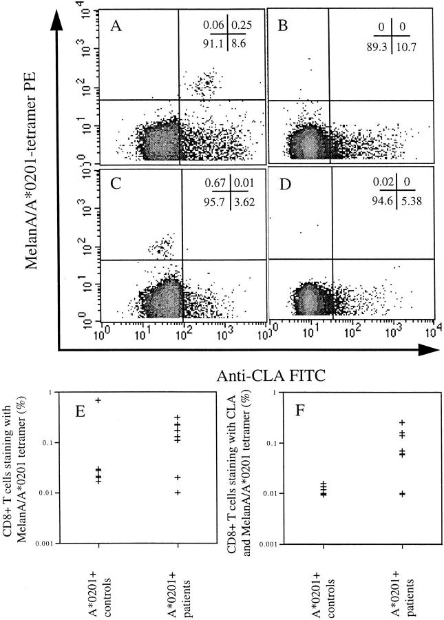
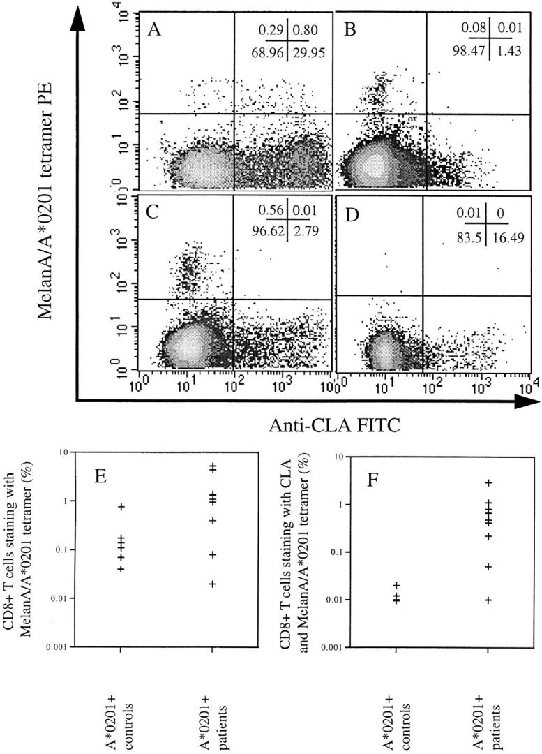
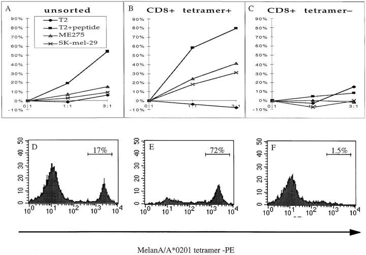
Vitiligo is an autoimmune condition characterized by loss of epidermal melanocytes. Using tetrameric complexes of human histocompatibility leukocyte antigen (HLA) class I to identify antigen-specific T cells ex vivo, we observed high frequencies of circulating MelanA-specific, A*0201-restricted cytotoxic T lymphocytes (A2-MelanA tetramer+ CTLs) in seven of nine HLA-A*0201-positive individuals with vitiligo. Isolated A2-MelanA tetramer+ CTLs were able to lyse A*0201-matched melanoma cells in vitro and their frequency ex vivo correlated with extent of disease. In contrast, no A2-MelanA tetramer+ CTL could be identified ex vivo in all four A*0201-negative vitiligo patients or five of six A*0201-positive asymptomatic controls. Finally, we observed that the A2-MelanA tetramer+ CTLs isolated from vitiligo patients expressed high levels of the skin homing receptor, cutaneous lymphocyte-associated antigen, which was absent from the CTLs seen in the single A*0201-positive normal control. These data are consistent with a role of skin-homing autoreactive melanocyte-specific CTLs in causing the destruction of melanocytes seen in autoimmune vitiligo. Lack of homing receptors on the surface of autoreactive CTLs could be a mechanism to control peripheral tolerance in vivo.


Figures

Figure 1
Figure 1
Uncultured CD8+ T cells stained with A2–MelanA tetramer and anti–CLA antigen from: (A) an A*0201-positive patient with vitiligo in whom the majority of MelanA-specific CTLs are positive for CLA (percentages given in each quadrant); (B) an A*0201-positive normal control individual in whom there were no detectable MelanA-specific CTLs ex vivo (pattern observed in five of six normal controls); (C) the A*0201- positive normal control individual in whom there were detectable MelanA-specific CTLs ex vivo, but which were all negative for CLA (pattern observed in one of six normal controls); (D) an A*0201-negative patient with vitiligo in whom there were no detectable A2–MelanA tetramer+ CTLs (observed in all A*0201-negative vitiligo patients). E shows the percentage of CD8+ T cells staining with A2–MelanA tetramer in the A*0201-positive normal controls and A*0201-positive vitiligo patients. F confirms a significant difference between the percentage of CLA-positive MelanA-specific CTLs in the PBMC of A*0201-positive patients and controls (P < 0.05).
Figure 2
Figure 2
Optimized peptide-stimulated IL-7–based cultures showing the CD8+ T cells stained with A2–MelanA tetramer and anti–CLA from: (A) an A*0201-positive patient with vitiligo in whom the majority of cultured MelanA-specific CTLs are positive for CLA (percentages given in each quadrant); (B) an A*0201-positive normal control individual in whom there was a low frequency of cultured MelanA-specific CTLs that were predominantly negative for CLA (pattern observed in five of six normal controls); (C) the A*0201-positive normal control individual in whom there were detectable MelanA-specific CTLs ex vivo, and in whom it was possible to culture high levels of MelanA-specific CTLs that were all persistently negative for CLA (pattern observed in one of six normal controls); (D) an A*0201-negative patient with vitiligo in whom there were no detectable A2–MelanA tetramer+ CTLs (observed in all A*0201-negative vitiligo patients) after a period of stimulation in vitro. E shows the percentage of CD8+ T cells from the cultures staining with A2–MelanA tetramer in the A*0201-positive normal controls and A*0201-positive vitiligo patients. F shows a significant difference between the percentage of CLA-positive MelanA-specific CTLs in the cultures from A*0201-positive patients and controls (P < 0.05).
Figure 3
Figure 3
(A–C) Cytolytic activity of MelanA/A*0201-specific CTL lines derived from an A*0201-positive vitiligo patient either unsorted (A), enriched for CD8+ A2–MelanA tetramer+ cells (B) or enriched for CD8+ A2–MelanA tetramer cells (C). The unsorted and CD8+-tetramer+ cells were both able to lyse peptide-pulsed targets, but only the CD8+ A2–MelanA tetramer+ population showed significant lysis of A*0201-matched melanoma cells (ME 275 and SK-mel-29). (D–E) The corresponding percentages of CD8+ T cells staining with the A2–MelanA tetramer are shown: the unsorted population contained 17% antigen-specific cells (D); the CD8+ A2–MelanA tetramer+ population contained 72% antigen-specific cells (E); and the CD8+ A2–MelanA tetramer population contained 1.5% antigen-specific cells.

References

    1. Song YH, Connor E, Li Y, Zorovich B, Balducci P, Maclaren N. The role of tyrosinase in autoimmune vitiligo. Lancet. 1994;344:1049–1052. - PubMed
    1. Kemp EH, Gawkrodger DJ, Watson PF, Weetman AP. Immunoprecipitation of melanogenic enzyme autoantigens with vitiligo sera: evidence for cross-reactive autoantibodies to tyrosinase and tyrosinase-related protein-2 (TRP-2) Clin Exp Immunol. 1997;109:495–500. - PMC - PubMed
    1. Hara I, Takechi Y, Houghton AN. Implicating a role for immune recognition of self in tumor rejection: passive immunization against the brown locus protein. J Exp Med. 1995;182:1609–1614. - PMC - PubMed
    1. Merimsky O, Shoenfeld Y, Baharav E, Altomonte M, Chaitchik S, Maio M, Ferrone S, Fishman P. Melanoma-associated hypopigmentation: where are the antibodies? . Am J Clin Oncol. 1996;19:613–618. - PubMed
    1. Naughton GK, Eisinger M, Bystryn JC. Antibodies to normal human melanocytes in vitiligo. J Exp Med. 1983;158:246–251. - PMC - PubMed

MeSH terms