. 1998 Sep 15;12(18):2887-98.
doi: 10.1101/gad.12.18.2887.

Repression of yeast Ste12 transcription factor by direct binding of unphosphorylated Kss1 MAPK and its regulation by the Ste7 MEK

Affiliations

Repression of yeast Ste12 transcription factor by direct binding of unphosphorylated Kss1 MAPK and its regulation by the Ste7 MEK

L Bardwell et al. Genes Dev. .

Abstract

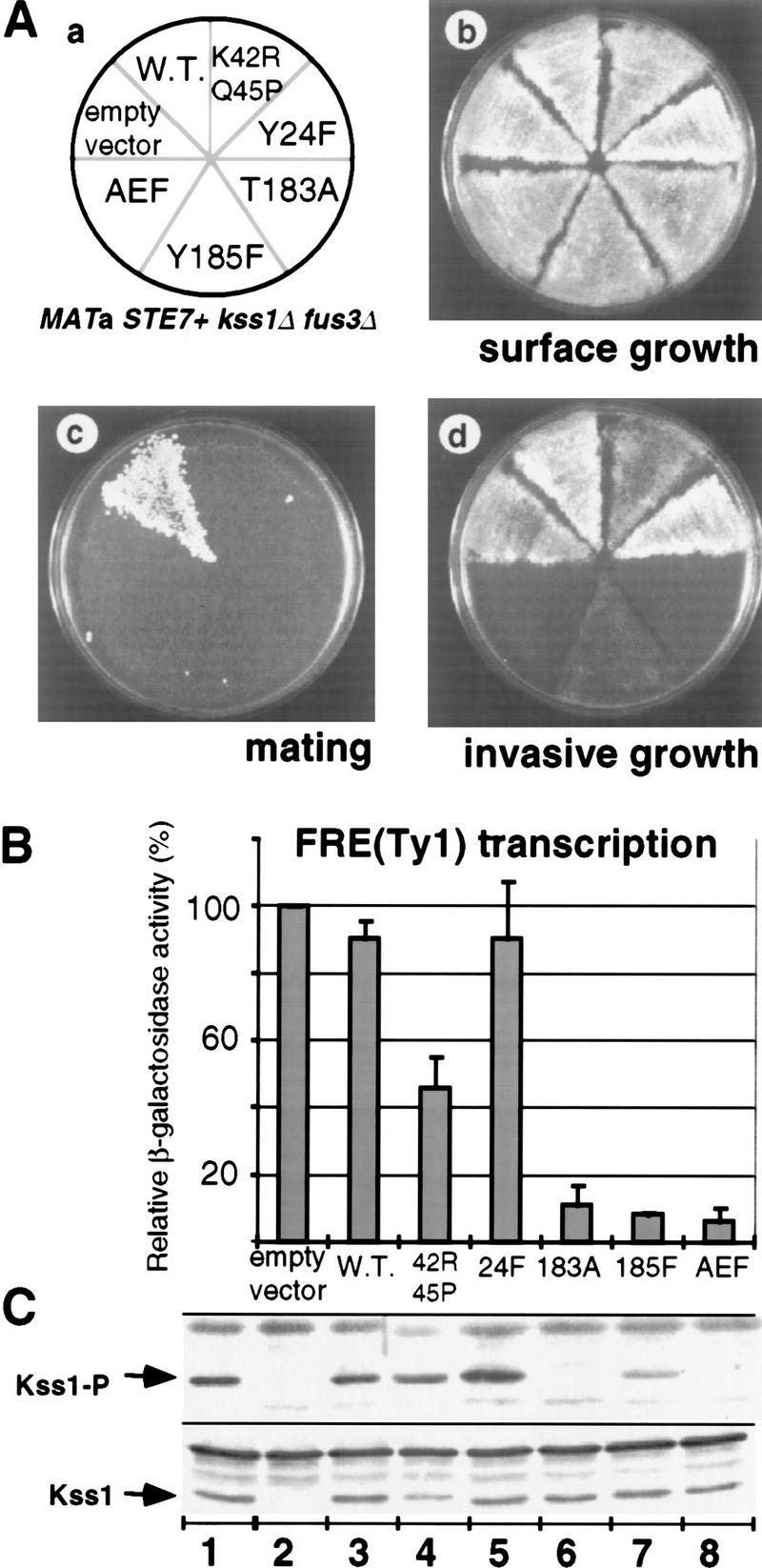
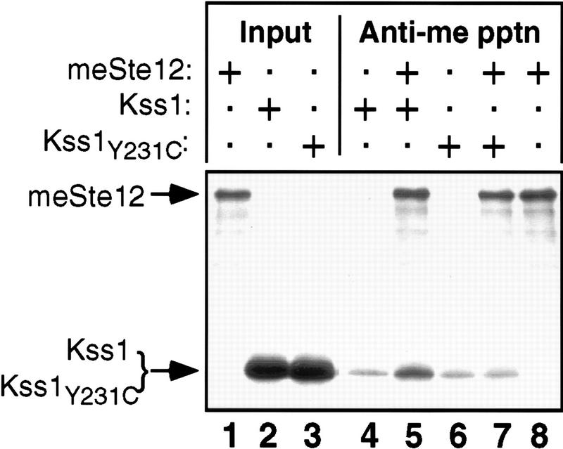
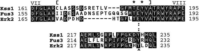
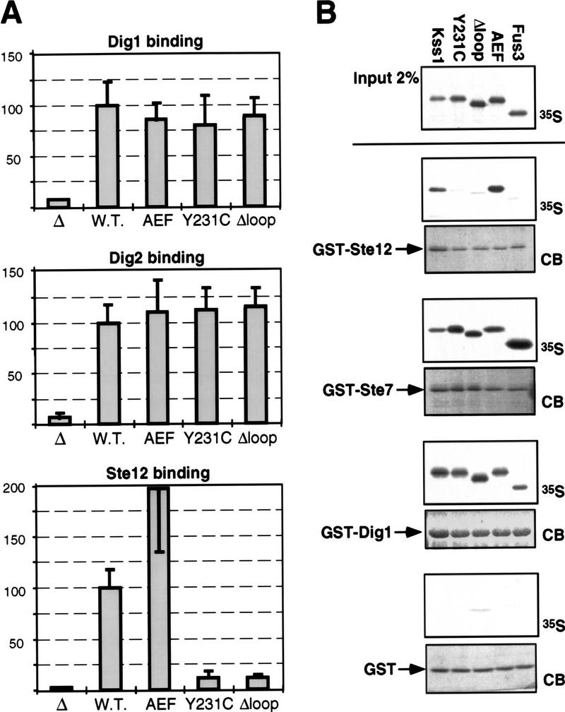
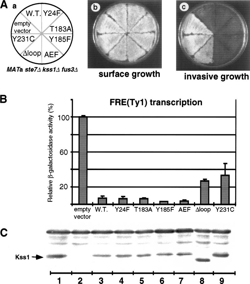
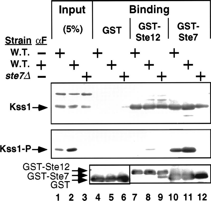
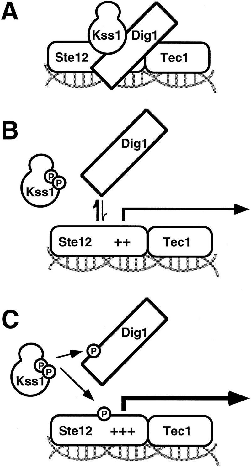
The mitogen-activated protein kinase (MAPK) Kss1 has a dual role in regulating filamentous (invasive) growth of the yeast Saccharomyces cerevisiae. The stimulatory function of Kss1 requires both its catalytic activity and its activation by the MAPK/ERK kinase (MEK) Ste7; in contrast, the inhibitory function of Kss1 requires neither. This study examines the mechanism by which Kss1 inhibits invasive growth, and how Ste7 action overcomes this inhibition. We found that unphosphorylated Kss1 binds directly to the transcription factor Ste12, that this binding is necessary for Kss1-mediated repression of Ste12, and that Ste7-mediated phosphorylation of Kss1 weakens Kss1-Ste12 interaction and relieves Kss1-mediated repression. Relative to Kss1, the MAPK Fus3 binds less strongly to Ste12 and is correspondingly a weaker inhibitor of invasive growth. Analysis of Kss1 mutants indicated that the activation loop of Kss1 controls binding to Ste12. Potent repression of a transcription factor by its physical interaction with the unactivated isoform of a protein kinase, and relief of this repression by activation of the kinase, is a novel mechanism for signal-dependent regulation of gene expression.


Figures

Figure 1
Figure 1
Phosphorylation of Kss1 is necessary and sufficient to permit haploid invasive growth. (A) Strain JCY130 (MATa STE7+ kss1Δ fus3Δ) was transformed with centromeric (low-copy) plasmids YCpU (empty vector), YCpU–KSS1 (W.T.), YCpU–kss1(K42R Q45P), YCpU–kss1(Y24F), YCpU–kss1(T183A), YCpU–kss1(Y185F), or YCpU–kss1(AEF) (a). The resulting transformants were streaked onto a plate selective for plasmid maintenance. After 2 days at 30°C, plates were replica-plated onto rich medium (YPD) plates and scored for surface growth (b), mating to strain DC17 (MATα his1) (c), or invasive growth after 30 hr at 30°C (d). (B) Expression of the FRETy1lacZ reporter gene. The strains described in A were transformed with plasmid YEpL–FTyZ, grown to mid-log phase in liquid medium, and β-galactosidase-specific activity was measured. Values are normalized to that observed for JCY130 carrying YCpU (3936 nmoles/min per milligram of protein) and represent the average of measurements, made in duplicate, on protein extracts prepared from at least three independent transformants of each strain (error bars indicate standard deviation). (C) Portions (20 μg) of the protein extracts used in B, plus an additional extract (lane 1) prepared from strain JCY100 (MATa KSS1+), were resolved on a 10% SDS–polyacrylamide gel and analyzed by immunoblotting with either anti-Kss1 (bottom) or anti-phospho–MAPK (top) antiserum.
Figure 2
Figure 2
Direct binding of Kss1 to Ste12 in vitro. Radiolabeled Kss1, Kss1Y231C, and myc epitope-tagged Ste12 (meSte12) were prepared by in vitro translation, partially purified by ammonium sulfate precipitation, and portions [for Kss1 and Kss1Y231C, 5% of the amount added in the immunoprecipitation (pptn) reactions, and for meSte12, 20%; Input] were resolved on a 10% SDS–polyacrylamide gel. Samples (2 pmoles) of Kss1 and Kss1Y231C, each accompanied by ∼200 μg of total protein from the rabbit reticulocyte lysate, were immunoprecipitated with the anti-c-Myc mAb 9E10 either in the absence (lanes 4,6) or presence (lanes 5,7,8) of 2 pmoles meSte12 protein, and the resulting immunoprecipitates were analyzed on the same gel. The percentage of the input Kss1 derivatives bound in the reactions corresponding to lanes 47 was, respectively, 0.6, 1.7, 0.5, and 0.7; and of meSte12 in lanes 5,7,8: 37.2, 30.7, and 35.1.
Figure 3
Figure 3
Structure and properties of the Kss1Δloop and Kss1Y231C derivatives. Amino acid sequence of Kss1 and homology to yeast Fus3 and rat Erk2 in the affected regions. Dashes (−) indicate single-residue gaps; identities are boxed. In Kss1Δloop protein, 21 residues of Kss1 (indicated by brackets) have been replaced with the unrelated 11-residue activation segment from S. cerevisiae Tpk3 (GenBank accession no. M17074), a homolog of the catalytic subunit of mammalian cyclic AMP-dependent protein kinase. Y231 is indicated by a carat (^). The target phosphoacceptor residues for MEK-mediated phosphorylation are indicated by asterisks (*). A hydrogen bond between Y231 and E184 in Erk2 is indicated by colons (:).
Figure 4
Figure 4
Kss1Δloop and Kss1Y231C are specifically defective in binding to Ste12. (A) Two-hybrid interaction of alleles of KSS1 with DIG1, DIG2, and STE12. Strain MaV103a was cotransformed with pKSS1–GDB (W.T.), pkss1(AEF)–GDB (encoding the T183A, Y185F derivative), pkss1(Y231C)–GDB, pkss1(Δloop)-GDB, or the plasmid encoding the GDB domain only (Δ); and with pGAD–DIG1(7–452), pGAD–DIG2(56–323), or pGAD–STE12(191–478); and β-galactosidase-specific activity was measured as described in the legend to Fig. 1B. Values are normalized to that observed for the interaction of wild-type KSS1 with DIG1, DIG2, or STE12 (41, 49 and 52 nmoles/min per milligram of protein, respectively). None of the KSS1–GDB alleles displayed an appreciable two-hybrid interaction with an empty GAD plasmid (data not shown). (B) In vitro binding of Kss1, Kss1 mutants, and Fus3 to Ste12, Ste7, and Dig1. Radiolabeled (35S) Kss1, Kss1Y231C, Kss1Δloop, Kss1AEF, and Fus3 proteins were prepared by in vitro translation, partially purified by ammonium sulfate precipitation, and portions (2% of the amount added in the binding reactions; Input) were resolved on a 10% SDS-polyacrylamide gel. Samples (1 pmole) of the same proteins were incubated with ∼2 μg of GST–Ste12298–473, GST–Ste71–172, GST–Dig1213–452, or GST bound to glutathione–Sepharose beads, and the resulting bead-bound protein complexes were isolated, resolved by 10% SDS-PAGE, and analyzed by staining with Coomassie blue (CB) for visualization of the bound GST fusion protein, and by autoradiography for visualization of the bound radiolabeled (35S) protein. GST, GST–Ste71–172, and GST–Dig1213–452 were purified from E. coli; GST–Ste12298–473 was purified from yeast (see Materials and Methods for details). The percentage of input Kss1, Kss1Y231C, Kss1Δloop, Kss1AEF, and Fus3 proteins bound to GST–Ste12 was, respectively: 1.6, 0.1, 0.1, 2.2, and 0.1; to GST–Ste7: 8.3, 13.6, 4.4, 6.1, and 30.4; to GST–Dig1: 9.1, 7.8, 7.4, 7.2, and 5.1; and to GST: 0.1 for all five proteins.
Figure 5
Figure 5
Ste12 binding contributes to Kss1-mediated inhibition of invasive growth. (A) Strain JCY137 (MATa ste7Δ kss1Δ fus3Δ) was transformed with plasmid YCpU (empty vector), YCpU–KSS1 (W.T.), YCpU–kss1(Y24F), YCpU–kss1(T183A), YCpU–kss1(Y185F), YCpU–kss1(AEF), YCpU–kss1(Δloop), or YCpU–kss1(Y231C) (a). The resulting transformants were streaked onto a plate selective for plasmid maintenance. After 2 days at 30°C, plates were replica-plated onto rich medium (YPD) plates and scored for surface growth (b), or invasive growth after 30 hr at 30°C (c). (B) Expression of the FRETy1–lacZ reporter gene. The strains described in A were transformed with plasmid YEpL–FTyZ, grown to mid-exponential phase in liquid medium, and β-galactosidase specific activity was measured as detailed in the legend to Fig. 1. Values are normalized to that observed for JCY137 carrying YCpU (3450 nmoles/min per milligram of protein). (C) Portions (20 μg) of the protein extracts used in B, plus an additional extract (lane 1) prepared from strain JCY100 (MATa KSS1+), were resolved on a 10% SDS-polyacrylamide gel and analyzed by immunoblotting with anti-Kss1 antiserum.
Figure 6
Figure 6
Phosphorylation of Kss1 reduces its affinity for Ste12. Cultures of strain JCY100 (W.T.) or JCY107 (ste7Δ), expressing Kss1 from a multicopy plasmid (YEpT–KSS1), were grown, treated (+), or not (−) with α-factor (αF) mating pheromone as indicated, and harvested. Cell extracts were prepared, and portions (5% of the amount added in the binding reactions; input) were resolved on a 10% SDS–polyacrylamide gel. Samples (1 mg) of the same extracts were incubated with ∼2 μg of GST–Ste12298–473, GST–Ste71–172, or GST bound to glutathione–Sepharose beads, and the resulting bead-bound protein complexes were isolated, resolved on the same gel, and analyzed by immunoblot analysis with anti-phospho–MAPK antiserum (middle) followed by stripping and reprobing with anti-Kss1 (top) and anti-GST (bottom) antiserum.
Figure 7
Figure 7
Model for Kss1-mediated regulation of Ste12 in invasive growth. (A) Unphosphorylated Kss1 binds directly to Ste12, and to Dig1 and Dig2, thereby stabilizing Dig1/2–Ste12 complexes and potentiating Dig-mediated repression of Ste12. (B) Phosphorylation of Kss1 by Ste7, in response to upstream signals, causes a conformational change in Kss1 that weakens its association with Ste12, and consequently reduces Dig1/2 interaction with Ste12–Tec1. (C) Phosphorylated and activated Kss1 further reinforces the transition, presumably by phosphorylating Dig1/2, Ste12, and/or Tec1, permitting full derepression.

References

    1. Bardwell L, Thorner J. A conserved motif at the amino termini of MEKs might mediate high-affinity interaction with the cognate MAPKs. Trends Biochem Sci. 1996;1:373–374. - PubMed
    1. Bardwell L, Cooper AJ, Friedberg EC. Stable and specific association between the yeast recombination and DNA repair proteins RAD1 and RAD10 in vitro. Mol Cell Biol. 1992;12:3041–3049. - PMC - PubMed
    1. Bardwell AJ, Bardwell L, Iyer N, Svejstrup JQ, Feaver WJ, Kornberg RD, Friedberg EC. Yeast nucleotide excision repair proteins Rad2 and Rad4 interact with RNA polymerase II basal transcription factor b (TFIIH) Mol Cell Biol. 1994a;14:3569–3576. - PMC - PubMed
    1. Bardwell L, Cook JG, Inouye CJ, Thorner J. Signal propagation and regulation in the mating pheromone response pathway of the yeast Saccharomyces cerevisiae. Devel Biol. 1994b;166:363–379. - PubMed
    1. Bardwell L, Cook JG, Chang EC, Cairns BR, Thorner J. Signaling in the yeast pheromone response pathway: Specific and high-affinity interaction of the mitogen-activated protein (MAP) kinases Kss1 and Fus3 with the upstream MAP kinase kinase Ste7. Mol Cell Biol. 1996;16:3637–3650. - PMC - PubMed

Publication types

MeSH terms

Substances