Comparative Study
. 1998 Sep 15;12(18):2932-42.
doi: 10.1101/gad.12.18.2932.

mei-W68 in Drosophila melanogaster encodes a Spo11 homolog: evidence that the mechanism for initiating meiotic recombination is conserved

Affiliations
Comparative Study

mei-W68 in Drosophila melanogaster encodes a Spo11 homolog: evidence that the mechanism for initiating meiotic recombination is conserved

K S McKim et al. Genes Dev. .

Abstract

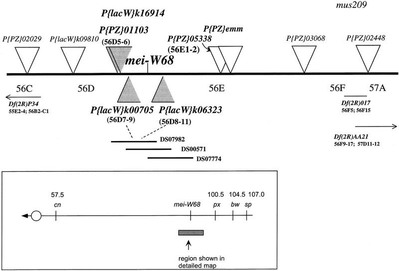
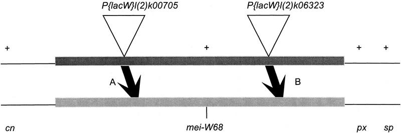
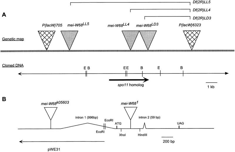
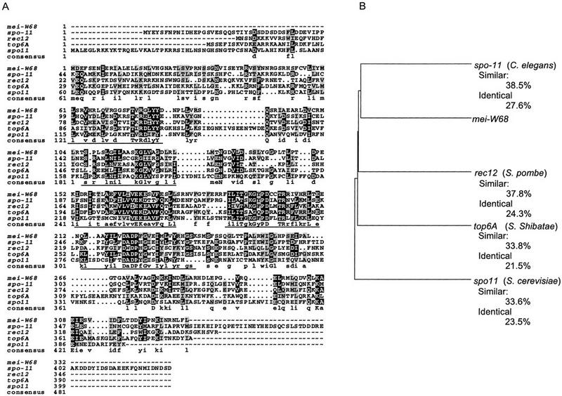
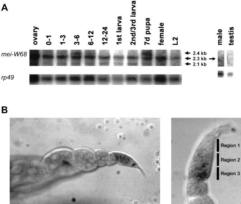
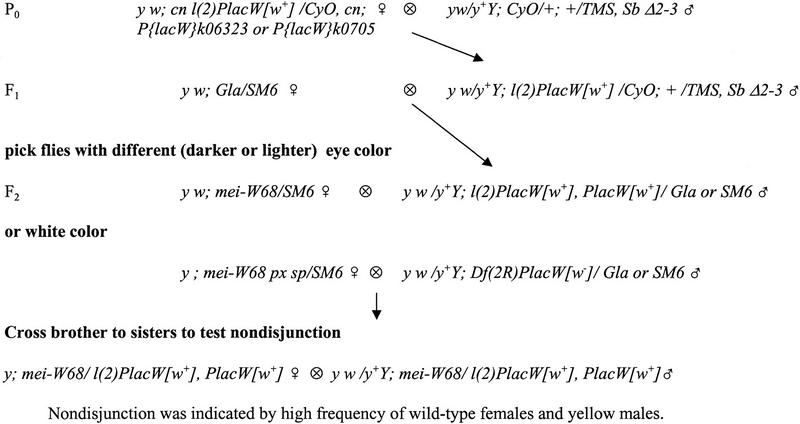
Meiotic recombination requires the action of several gene products in both Saccharomyces cerevisiae and Drosophila melanogaster. Genetic studies in D. melanogaster have shown that the mei-W68 gene is required for all meiotic gene conversion and crossing-over. We cloned mei-W68 using a new genetic mapping method in which P elements are used to promote crossing-over at their insertion sites. This resulted in the high-resolution mapping of mei-W68 to a <18-kb region that contains a homolog of the S. cerevisiae spo11 gene. Molecular analysis of several mutants confirmed that mei-W68 encodes an spo11 homolog. Spo11 and MEI-W68 are members of a family of proteins similar to a novel type II topoisomerase. On the basis of this and other lines of evidence, Spo11 has been proposed to be the enzymatic activity that creates the double-strand breaks needed to initiate meiotic recombination. This raises the possibility that recombination in Drosophila is also initiated by double-strand breaks. Although these homologous genes are required absolutely for recombination in both species, their roles differ in other respects. In contrast to spo11, mei-W68 is not required for synaptonemal complex formation and does have a mitotic role.


Figures

Figure 1
Figure 1
Genetic map of the mei-W68 region. mei-W68 was mapped relative to the P-element insertions (triangles). The P-element insertions flanking the mei-W68 locus that were inserted within the overlap of P1 clones DS07982 and DS00571 are shaded. Deficiencies that complemented mei-W68 are shown with their cytogenetic breakpoints. The P{PZ}05338 insertion is an allele of smooth (Lage et al. 1997). P{PZ}01103 is inserted at the hts locus (Berkeley Drosphila Genome Project).
Figure 2
Figure 2
The types of recombinants recovered from P element-mediated male crossing-over. This schematic map shows the markers used in most of the mapping experiments. It is not drawn to scale as the two insertions are much closer together relative to the flanking markers cn, px, and sp. If the P element was to the left (distal) of mei-W68, the + px sp crossovers (A) also carried the mei-W68 mutation. Conversely, if the insertion was to the right (proximal) of mei-W68, then the + px sp cross overs (B) did not carry the mei-W68 mutation. Shown is the direction of only one of the two crossover classes that was recovered from each experiment. Relative to the + px sp crossovers, the cn + + crossovers had the opposite linkage arrangement with mei-W68.
Figure 3
Figure 3
Physical map of the mei-W68 region. (A) The restriction map came from two sources: our own restriction mapping of DS00571, and the Genome Project sequencing of the overlapping P1 DS07982. The molecular structures of three mei-W68 deletion mutants are shown. The brackets above the genetic map are to indicate that the LL4, LL5, and LD3 chromosomes are deletions with one endpoint at the original P{lacW}l(2)k06323 insertion site. (B) Depiction of the mei-W68 transcript showing the intron/exon structure and the location of the insertions in mei-W681 and mei-W68k05603. To show that the mei-W681 insertion was not in the first or second introns, we probed HindIII and XhoI digests with labeled EcoRI fragment pWE31 (B). The appearance of the larger insertion band in the HindIII digest and the wild-type bands in the XhoI digest (data not shown) showed that the insertion site was between the first and second introns. The position of the k05603 insertion was determined from sequence obtained by the genome project off the 5′ end of the P element (Berkeley Drosphila Genome Project).
Figure 4
Figure 4
mei-W68 is a homolog of spo11 and the topo6A type II topoisomerase. (A) Protein sequence alignment spo11 homologs from S. cerevisiae Spo11 (Atcheson et al. 1987), Schizosaccharomyces pombe Rec12 (Lin and Smith 1994), Sulfolobus shibatae topo6A (Bergerat et al. 1997), and the predicted C. elegans gene from cosmid T05E11.4 (Wilson et al. 1994; Dernburg et al. 1998). (B) Dendogram comparing the extent of similarity between the different proteins. The percentages of similar and identical amino acids are relative to MEI-W68.
Figure 5
Figure 5
Developmental Northern blot using a mei-W68 antisense RNA probe. The ribosomal protein gene RP49 was used as a standard for the amount of mRNA loaded on the gel. (A) There are three transcripts of 2.4, 2.3, and 2.1 kb. The 2.3-kb transcript is seen through all developmental stages. The largest transcript (2.4 kb) is detected in the first 12 hr of embryogenesis and in the pupal stage. The 2.1-kb transcript is seen in all stages but is clear at 12–24 hr embryo stage and larval stages. A transcript was difficult to detect in males but was detected by RT–PCR (see text). The numbers for lanes 2–6 indicate the hours of embryonic development. (7d) The pupal stage at day 7 of development. (B) Two examples of an in situ hybridization to whole ovaries using an RNA probe made from the mei-W68 full-length cDNA. (Left) The transcript was detectable in the latter half of the germarium, which corresponds to regions 2 and 3, but there was little or no transcript was detectable in later stages. (Right) Another germarium at higher magnification. The transcript was not detected in region 1, where the premeiotic mitotic divisions occur.
Figure 6
Figure 6
Genetic crosses to generate P element-induced local-hop insertions and deletions of the mei-W68 gene. CyO and SM6 are second chromosome balancers carrying the dominant Cy maker; TMS is a third chromosome balancer carrying the source of P-element transposase, Δ2-3 and the dominant Sb marker. In the F1 generation, the P elements are exposed to transposase, and any mobilization events are detected in the F2 by a change in eye color. These exceptional flies were picked and mated separately to establish stocks and test for nondisjunction (Materials and Methods).

References

    1. Altschul SF, Gish W, Miller W, Meyers EW, Lipman DJ. Basic local alignment search tool. J Mol Biol. 1990;215:403–410. - PubMed
    1. Atcheson CL, DiDomenico B, Frackman S, Esposito RE, Elder RT. Isolation, DNA sequence, and regulation of a meiosis-specific eukaryotic recombination gene. Proc Natl Acad Sci. 1987;84:8035–8039. - PMC - PubMed
    1. Baker BS, Carpenter ATC. Genetic analysis of sex chromosomal meiotic mutants in Drosophila melanogaster. Genetics. 1972;71:255–286. - PMC - PubMed
    1. Baker BS, Carpenter ATC, Ripoll P. The utilization during mitotic cell division of loci controlling meiotic recombination in Drosophila melanogaster. Genetics. 1978;90:531–578. - PMC - PubMed
    1. Berger JM, Gamblin SJ, Harrison SC, Wang JC. Structure and mechanism of DNA topoisomerase II. Nature. 1996;379:225–232. - PubMed

Publication types

MeSH terms

Associated data