. 1998 Sep 21;142(6):1461-71.
doi: 10.1083/jcb.142.6.1461.

Progressive muscular dystrophy in alpha-sarcoglycan-deficient mice

Affiliations

Progressive muscular dystrophy in alpha-sarcoglycan-deficient mice

F Duclos et al. J Cell Biol. .

Abstract

Limb-girdle muscular dystrophy type 2D (LGMD 2D) is an autosomal recessive disorder caused by mutations in the alpha-sarcoglycan gene. To determine how alpha-sarcoglycan deficiency leads to muscle fiber degeneration, we generated and analyzed alpha-sarcoglycan- deficient mice. Sgca-null mice developed progressive muscular dystrophy and, in contrast to other animal models for muscular dystrophy, showed ongoing muscle necrosis with age, a hallmark of the human disease. Sgca-null mice also revealed loss of sarcolemmal integrity, elevated serum levels of muscle enzymes, increased muscle masses, and changes in the generation of absolute force. Molecular analysis of Sgca-null mice demonstrated that the absence of alpha-sarcoglycan resulted in the complete loss of the sarcoglycan complex, sarcospan, and a disruption of alpha-dystroglycan association with membranes. In contrast, no change in the expression of epsilon-sarcoglycan (alpha-sarcoglycan homologue) was observed. Recombinant alpha-sarcoglycan adenovirus injection into Sgca-deficient muscles restored the sarcoglycan complex and sarcospan to the membrane. We propose that the sarcoglycan-sarcospan complex is requisite for stable association of alpha-dystroglycan with the sarcolemma. The Sgca-deficient mice will be a valuable model for elucidating the pathogenesis of sarcoglycan deficient limb-girdle muscular dystrophies and for the development of therapeutic strategies for this disease.


Figures

Figure 1
Figure 1
Generation of Sgca-null mutant mice. (a) Restriction map of the wild-type Sgca locus (Sgca +), the targeting construct, and the targeted locus. A region of 902 bp including exons 2 and 3 (E2 and E3) was deleted and replaced by a phosphoglycerate kinase-neomycin cassette (NEOR). (b) Southern blot analysis. Using the probe shown (black box), the targeted locus contains an EcoR1 fragment of 8.8 kb, whereas the intact allele shows a 5.5-kb band: clone 594 is correctly targeted. (c) Genotyping by PCR. Primer sites are shown in a; using primers INT1 and EX2 the wild-type allele (+/+) corresponds to a 1,061-bp fragment; using primers INT1 and NEOTR, the null allele is 618 bp. (d) Northern blotting. An α-sarcoglycan cDNA probe reveals the correct sized transcript in wild type (+/+) and heterozygotes (+/−) from 30 μg total RNA extracted from skeletal muscle; homozygous mutant tissue shows no α-sarcoglycan transcript. (e) Western blot analysis. Using an affinity-purified polyclonal antibody against a COOH-terminal peptide of α-sarcoglycan, membrane-enriched preparations of skeletal muscle reveal the protein in (+/+), and (+/−), but not in (−/−).
Figure 6
Figure 6
Abnormal contractile properties of Sgca-deficient muscle. The data on EDL and soleus muscles of the Sgca-null mutant mice are represented as a percentage of the values for muscles of control mice. The diagram shows bar graphs for the data on body mass, muscle mass, absolute maximum isometric tetanic force, specific force, and peak force during resistance to stretch of passive muscles. Asterisk, significant differences between the data obtained in Sgca-deficient and control mice. All data are presented as the mean ± one SEM.
Figure 4
Figure 4
Immunofluores- cence analysis of sarcolemma proteins in Sgca-deficient skeletal and cardiac muscle. Skeletal (a) and cardiac (b) muscle cryosections from wild-type (+/+) and Sgca-null (−/−) mice were stained with antibodies against dystrophin (DYS), α-dystroglycan (α-DG), β-dystroglycan (β-DG), laminin α2-chain, (lamα2), α-, β-, γ-, δ-, and ε-sarcoglycan (SG), and sarcospan (SPN). The sarcoglycans and sarcospan were drastically reduced in the Sgca-deficient muscle whereas the α-sarcoglycan homologue ε-sarcoglycan was present in comparable amount to control. Dystrophin staining is reduced in Sgca-null mutant mice from the sarcolemma of skeletal muscle, whereas it is maintained at equal levels to control in cardiomyocytes. Bar, 50 μm.
Figure 4
Figure 4
Immunofluores- cence analysis of sarcolemma proteins in Sgca-deficient skeletal and cardiac muscle. Skeletal (a) and cardiac (b) muscle cryosections from wild-type (+/+) and Sgca-null (−/−) mice were stained with antibodies against dystrophin (DYS), α-dystroglycan (α-DG), β-dystroglycan (β-DG), laminin α2-chain, (lamα2), α-, β-, γ-, δ-, and ε-sarcoglycan (SG), and sarcospan (SPN). The sarcoglycans and sarcospan were drastically reduced in the Sgca-deficient muscle whereas the α-sarcoglycan homologue ε-sarcoglycan was present in comparable amount to control. Dystrophin staining is reduced in Sgca-null mutant mice from the sarcolemma of skeletal muscle, whereas it is maintained at equal levels to control in cardiomyocytes. Bar, 50 μm.
Figure 5
Figure 5
Immunoblot analysis of skeletal muscle membranes. Skeletal muscle microsomes from control (+/+) and Sgca-deficient (−/−) mice were analyzed by 3–15% SDS-PAGE and immunoblotting using antibodies against several DGC components. In particular we used antibodies against the sarcoglycans (α-, β-, γ-, δ-, and ε-SG), dystroglycans (α- and β-DG), and dystrophin (DYS). In addition we stained blots with antibodies against neuronal nitric oxide synthase (NOS), which has been shown to be associated with dystrophin, and the dystrophin homologue utrophin (UTR). To demonstrate equal loading of protein samples we used the α1 subunit of the dihydropyridine receptor (α1-DHPR) and caveolin-3 (CAV-3) as positive markers.
Figure 2
Figure 2
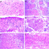
Histological analysis of Sgca-deficient diaphragm muscle. Sgca-null mutant mice started to develop a progressive muscular dystrophy at 1 wk of age and, in contrast to mdx mice, showed ongoing muscle necrosis with increasing age. Examples of muscle pathology in the diaphragm of different aged mice are demonstrated. (a) Myocyte atrophy (small fibers scattered throughout the micrograph) and central nucleation (see fibers near the center and in upper left quadrant) after regeneration in an 8-d-old mouse. (b) A small focus of myocyte necrosis (center) with surrounding regenerating and atrophic fibers in an 18-d-old mouse. (c) Skeletal muscle of a 4-wk-old mouse with ongoing necrosis (right of center), regeneration, central nucleation, endomysial fibrosis (increase in tissue between muscle fibers), atrophy, hypertrophy (large fibers in left lower quadrant), and fiber splitting. (d) The edge of a large, confluent area of acute myocyte necrosis in an 8-wk-old mouse. (e) More severe endomysial fibrosis with atrophy, central nucleation, fiber splitting, and dystrophic calcification (dark structures in lower right) in an 8-wk-old mouse. (f) Small foci of myocyte necrosis surrounded by atrophic and hypertrophic fibers, central nucleation, and fiber splitting are shown in a 16-wk-old mouse. All panels show 7-μm frozen sections of diaphragm muscle stained with H & E.
Figure 3
Figure 3
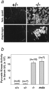
Evaluation of sarcolemma permeability. (a) Heterozygous (+/−) and homozygous-null (−/−) mice were intravenously injected with EBD. The panels show dye uptake into muscle fibers of the femoral quadriceps and diaphragm muscles 6 h after injection. Dye accumulation was only detected in skeletal muscle from Sgca-null mutants. Activity of muscle-specific PK in 7–10-wk-old wild-type (+/+), heterozygotes (+/−), homozygotes (−/), and mdx mice (b). Measurement of PK released from the muscle fiber into the circulating blood showed similar high levels of PK activity in (−/−) and mdx mice compared with (+/−) and control (+/+). Error bars indicate the standard deviation where n equals the number of mice in each set. Bar, 50 μm.
Figure 7
Figure 7
Restoration of sarcoglycan complex after adenovirus injection. Recombinant α-sarcoglycan adenovirus mediates restoration of DGC components to the sarcolemma. 2-d-old Sgca-null mutant pups were injected in their hamstring muscle with a recombinant α-sarcoglycan adenovirus containing the human α-sarcoglycan coding sequence under the control of a viral RSV promoter. Serial transverse cryosections of injected muscle after 3 wk, were stained with antibodies against α-sarcoglycan (a), β-sarcoglycan (b), γ-sarcoglycan (c), and δ-sarcoglycan (d), dystrophin (e), and sarcospan (f). Bar, 50 μm.

References

    1. Allamand V, Sunada Y, Salih MA, Straub V, Ozo CO, Al-Turaiki MH, Akbar M, Kolo T, Colognato H, Zhang X, Sorokin LM, Yurchenco PD, Tryggvason K, Campbell KP. Mild congenital muscular dystrophy in two patients with an internally deleted laminin alpha2-chain. Hum Mol Genet. 1997;6:747–752. - PubMed
    1. Bönnemann CG, Modi R, Noguchi S, Mizuno Y, Yoshida M, Gussoni E, McNally EM, Duggan DJ, Angelini C, et al. Beta-sarcoglycan (A3b) mutations cause autosomal recessive muscular dystrophy with loss of the sarcoglycan complex. Nat Genet. 1995;11:266–273. - PubMed
    1. Brenman JE, Chao DS, Xia H, Aldape K, Bredt DS. Nitric oxide synthase complexed with dystrophin and absent from skeletal muscle sarcolemma in Duchenne muscular dystrophy. Cell. 1995;82:743–752. - PubMed
    1. Brooks SV, Faulkner JA. Contractile properties of skeletal muscles from young, adult and aged mice. J Physiol (Lond) 1988;404:71–82. - PMC - PubMed
    1. Carrie A, Piccolo F, Leturcq F, de Toma C, Azibi K, Beldjord C, Vallat JM, Merlini L, Voit T, Sewry C, Urtizberea JA, Romero N, Tome FM, Fardeau M, Sunada Y, Campbell KP, Kaplan JC, Jeanpierre M. Mutational diversity and hot spots in the alpha-sarcoglycan gene in autosomal recessive muscular dystrophy (LGMD2D) J Med Genet. 1997;34:470–475. - PMC - PubMed

Publication types

MeSH terms

Associated data