. 1998 Sep 21;142(6):1533-45.
doi: 10.1083/jcb.142.6.1533.

Activation of the MKK/ERK pathway during somatic cell mitosis: direct interactions of active ERK with kinetochores and regulation of the mitotic 3F3/2 phosphoantigen

Affiliations

Activation of the MKK/ERK pathway during somatic cell mitosis: direct interactions of active ERK with kinetochores and regulation of the mitotic 3F3/2 phosphoantigen

P S Shapiro et al. J Cell Biol. .

Abstract

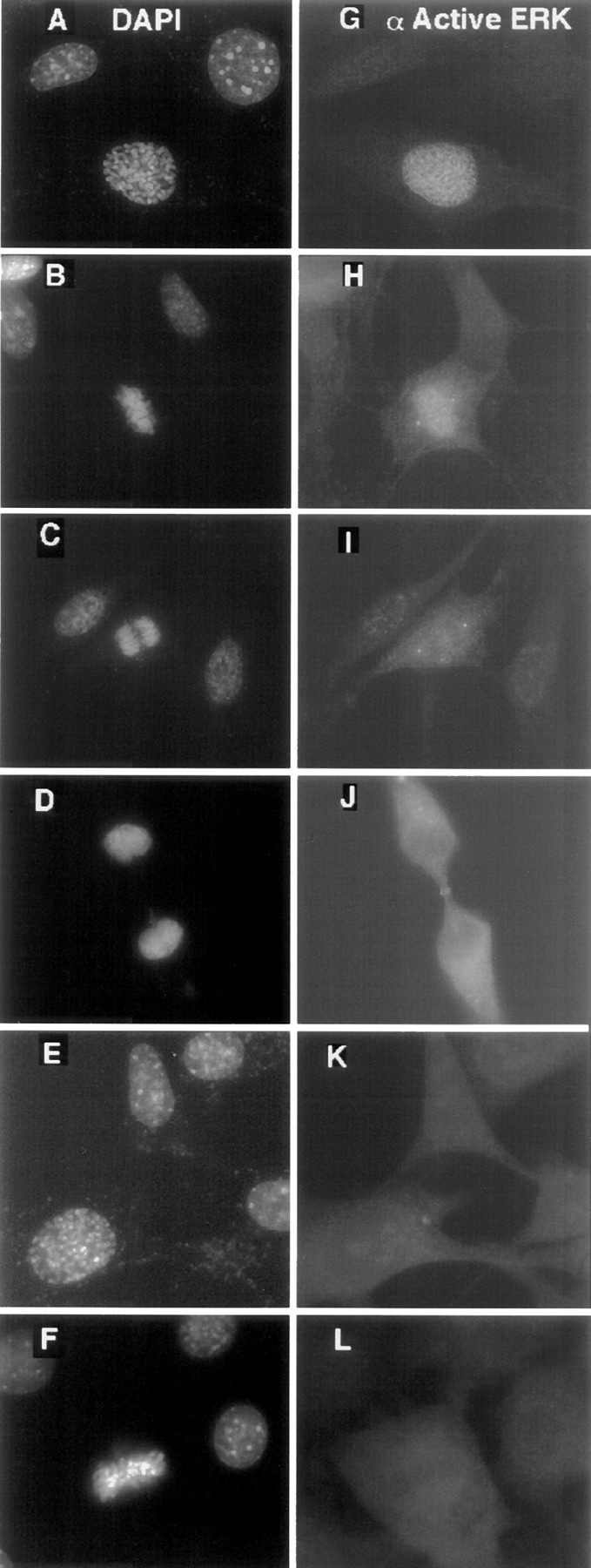
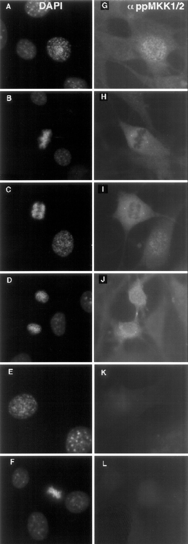
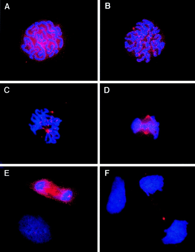
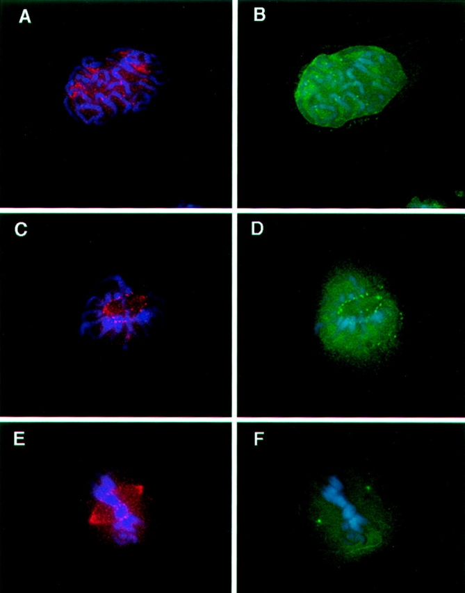
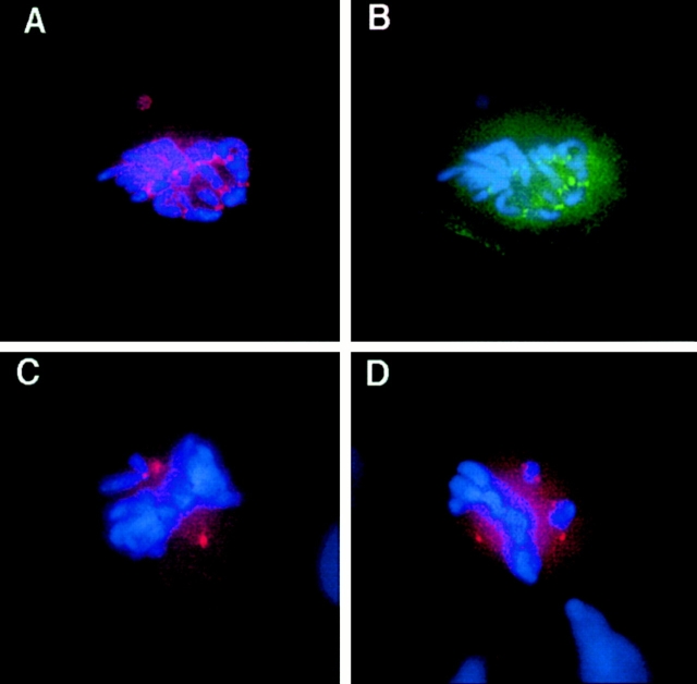
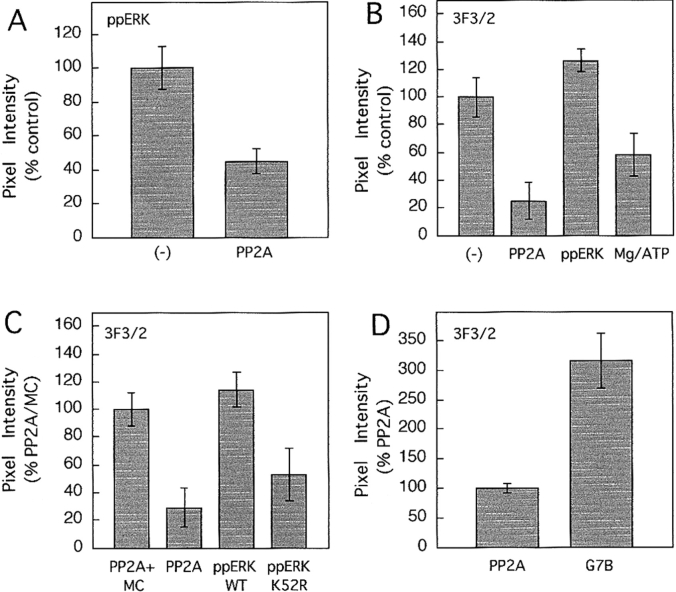
The mitogen-activated protein (MAP) kinase pathway, which includes extracellular signal-regulated protein kinases 1 and 2 (ERK1, ERK2) and MAP kinase kinases 1 and 2 (MKK1, MKK2), is well-known to be required for cell cycle progression from G1 to S phase, but its role in somatic cell mitosis has not been clearly established. We have examined the regulation of ERK and MKK in mammalian cells during mitosis using antibodies selective for active phosphorylated forms of these enzymes. In NIH 3T3 cells, both ERK and MKK are activated within the nucleus during early prophase; they localize to spindle poles between prophase and anaphase, and to the midbody during cytokinesis. During metaphase, active ERK is localized in the chromosome periphery, in contrast to active MKK, which shows clear chromosome exclusion. Prophase activation and spindle pole localization of active ERK and MKK are also observed in PtK1 cells. Discrete localization of active ERK at kinetochores is apparent by early prophase and during prometaphase with decreased staining on chromosomes aligned at the metaphase plate. The kinetochores of chromosomes displaced from the metaphase plate, or in microtubule-disrupted cells, still react strongly with the active ERK antibody. This pattern resembles that reported for the 3F3/2 monoclonal antibody, which recognizes a phosphoepitope that disappears with kinetochore attachment to the spindles, and has been implicated in the mitotic checkpoint for anaphase onset (Gorbsky and Ricketts, 1993. J. Cell Biol. 122:1311-1321). The 3F3/2 reactivity of kinetochores on isolated chromosomes decreases after dephosphorylation with protein phosphatase, and then increases after subsequent phosphorylation by purified active ERK or active MKK. These results suggest that the MAP kinase pathway has multiple functions during mitosis, helping to promote mitotic entry as well as targeting proteins that mediate mitotic progression in response to kinetochore attachment.


Figures

Figure 1
Figure 1
Specificity of anti-ACTIVE MAPK antibody. (A) Immunoblots of wild type or mutant recombinant ERK2 phosphorylated with constitutively active mutant MKK1-G1C. (Top) Immunoreactivity with anti-ACTIVE MAPK antibody. (Bottom) Reactivity with antibody recognizing the COOH terminus of ERK2, where ERK2 gel mobility is retarded upon phosphorylation. (B) Immunoblots of whole cell extracts (20 μg) from NIH 3T3, CHO, and PtK1 cells that were serum-depleted overnight, and then treated with or without 10% serum + 0.1 μM PMA for 5 min (PMA). (Top) Specific immunoreactivity of anti-ACTIVE MAPK antibody with phosphorylated ERK1 and ERK2 (arrows). Only phosphorylated ERK2 was observed in CHO cell lysates. (Bottom) Corresponding levels of ERK2, probed with an antibody recognizing the COOH terminus of ERK2. Filled arrows show phosphorylated ERK2 with retarded mobility, and open arrows show unphosphorylated ERK2.
Figure 2
Figure 2
Mitotic activation of ERK in NIH 3T3 cells. Cells were fixed with glutaraldehyde, and were then stained with DAPI (A– F ) and anti-ACTIVE MAPK antibody probed with Texas Red– conjugated secondary antibody (G–L). Fluorescence of representative cells in (A and G) interphase (upper cells) and prophase (lower cell); (B and H) metaphase; (C and I) anaphase; and (D and J) telophase are shown. Note active ERK staining at spindle poles in metaphase and anaphase (H and I). Staining patterns were reproduced with paraformaldehyde or NBF/methanol fixation. (E, F, K, and L) In peptide competition controls, anti-ACTIVE MAPK antibody was preincubated with 0.1 mg/ml phosphopeptide before incubation with coverslips. Under these conditions, mitotic cells showed no ERK immunoreactivity, as demonstrated with prophase (E and F) and metaphase (K and L) cells.
Figure 3
Figure 3
Activation of ERK during prophase precedes nuclear envelope breakdown. NIH 3T3 cells were fixed with glutaraldehyde, and were then stained with DAPI (A and D) and costained with (B and E) anti-ACTIVE MAPK antibodies probed with Texas Red–coupled secondary antibodies or (C and F) lamin B antibodies probed with fluorescein isothiocyanate-conjugated secondary antibodies. Patterns of lamin B staining indicate that nuclear envelope membranes are intact in prophase cells during ERK activation. Corresponding interphase cells are shown to the left of each prophase cell in the two representative images.
Figure 4
Figure 4
Specificity of anti-phosphoMEK1/2 antibody. (A) Immunoblots of wild-type or mutant recombinant MKK1, after phosphorylation with constitutively active mutant MEKKΔC. (Top) Immunoreactivity with anti-phosphoMEK1/2 antibody. (Bottom) Reactivity with antibody recognizing the NH2 terminus of MKK1. Differences in gel mobility are a result of removing the 5-kD NH2 terminal (His)6 tag from mono- or diphosphorylated MKK1, and deleting residues 44–51 in MKK1-G1C. (B) Immunoblots of whole cell extracts (20 μg) from NIH 3T3, CHO, and PtK1 cells that were serum-depleted overnight, and then treated with or without 10% serum + 0.1 μM PMA for 30 min. (Top) Specific immunoreactivity of anti-phosphoMEK1/2 antibody with phosphorylated MKK1 and MKK2, indicated by arrows. (Bottom) Corresponding levels of MKK1 probed with an antibody recognizing the NH2 terminus of MKK1.
Figure 5
Figure 5
Mitotic activation of MKK in NIH 3T3 cells. Cells were fixed with NBF/methanol, and were then stained with DAPI (A– F) and anti-phosphoMEK1/2 antibody probed with Texas Red– conjugated secondary antibody (F–L). Fluorescence of representative cells in (A and G) interphase and prophase; (B and H) metaphase; (C and I) anaphase; and (D and J) telophase are shown. Staining patterns were reproduced with paraformaldehyde or glutaraldehyde fixation. (E, F, K, and L) In peptide competition controls, anti-phosphoMEK1/2 antibody was preincubated with 0.1 mg/ml phosphopeptide before incubation with coverslips. Under these conditions, mitotic cells showed no active MKK immunoreactivity as demonstrated with prophase (E and F) and metaphase (K and L) cells.
Figure 6
Figure 6
Mitotic activation of ERK in PtK1 cells. Cells were fixed with glutaraldehyde, and were then stained with DAPI and anti-ACTIVE MAPK antibody probed with Texas Red–conjugated secondary antibody. Fluorescence of representative cells in (A) early prophase; (B) late prophase; (C) prometaphase; (D) metaphase; (E) anaphase; and (F) telophase. Interphase cells are also shown in E and F. Staining patterns were also reproduced with paraformaldehyde or NBF/methanol fixation. Controls show no immunoreactivity when anti-ACTIVE MAPK antibody was preincubated with 0.1 mg/ml phosphopeptide before incubation with coverslips (data not shown).
Figure 7
Figure 7
Active ERK and CENP/E colocalize at kinetochores. PtK1 cells were fixed with glutaraldehyde, and were then stained with DAPI together with mouse monoclonal anti-phosphoMAPK antibody and rabbit anti-CENP-E antibody, probed with Texas Red– or fluorescein isothiocyanate–conjugated secondary antibodies, respectively. Fluorescence of representative cells in (A and B) prophase; (C and D) prometaphase; and (E and F) anaphase; showing kinetochore staining of active ERK early in prophase, before the appearance of CENP-E, and disappearance of kinetochore-associated active ERK during anaphase before the disappearance of CENP-E. An interphase cell is also shown in E and F.
Figure 8
Figure 8
Active ERK localized to kinetochores is a minor fraction of the total ERK pool. PtK1 cells were fixed with glutaraldehyde, and were then stained with (A) COOH-terminal ERK2 antibody probed with Texas Red–conjugated secondary antibody; or (B) DAPI. Fluorescence of representative cells in prophase and interphase are shown in each panel. Although a significant percentage of ERK is nuclear in prophase, kinetochore localization cannot be detected.
Figure 9
Figure 9
Mitotic activation of MKK in PtK1 cells. Cells were fixed with glutaraldehyde, and were then stained with DAPI and anti-phosphoMEK1/2 antibody probed with Texas Red–conjugated secondary antibody. Fluorescence of representative cells in (A) prophase; (B) prometaphase; (C) metaphase; and (D) anaphase. Staining patterns were reproduced with paraformaldehyde or NBF/methanol fixation. Controls show no immunoreactivity when anti-phosphoMEK1/2 antibody was preincubated with 0.1 mg/ml phosphopeptide before incubation with coverslips (data not shown).
Figure 10
Figure 10
Active ERK and 3F3/2 antigen colocalize at kinetochores. PtK1 cells were fixed with glutaraldehyde, and were then stained with DAPI, together with anti-ACTIVE MAPK antibody probed with Texas Red–coupled secondary antibody and 3F3/2 antibody probed with fluorescein isothiocyanate-conjugated secondary antibody. Fluorescence of representative cells in (A and B) prophase; (C and D) prometaphase; and (E and F) metaphase, showing corresponding appearances of active ERK and 3F3/2 antigen at kinetochores early in prophase, and disappearance at or after metaphase.
Figure 11
Figure 11
Kinetochore localization of active ERK correlates with microtubule disruption or chromosome displacement. (A and B) PtK1 cells were treated with nocodazole for 30 min, and were then fixed with glutaraldehyde and stained with DAPI, anti-ACTIVE MAPK antibody, and 3F3/2 antibody as in Fig. 10. Fluorescence of representative cells in metaphase showed stable kinetochore staining of (A) active ERK and (B) 3F3/2 antigen. (C and D) Untreated PtK1 cells fixed and stained with DAPI or anti-ACTIVE MAPK antibody. High immunoreactivity of active ERK is observed on metaphase chromosomes that are misaligned from the metaphase plate.
Figure 12
Figure 12
Kinetochore localization of active ERK and 3F3/2 antigen on isolated chromosomes. (A and B) Isolated CHO cell chromosomes bound to glass coverslips were stained without fixation with DAPI and (A) anti-ACTIVE MAPK antibody or (B) 3F3/2 antibody, probed in each case with Texas Red–conjugated secondary antibodies. (C and D) Isolated chromosomes were incubated with added (C) PP2A for 15 min, or (D) PP2A for 15 min, followed by 2 μM microcystin, 2 μg active ERK2, and MgATP for 10 min. Chromosomes were then stained with DAPI and 3F3/2 antibody, followed immediately by data collection.
Figure 13
Figure 13
Sensitivity of active ERK or 3F3/2 antigen at isolated kinetochores to dephosphorylation or rephosphorylation by added enzymes. (A) Isolated chromosome samples bound to glass coverslips were incubated without or with PP2A (2U) for 15 min, followed by staining with DAPI and anti-ACTIVE MAPK antibody, as in Fig. 12. 8–14 kinetochores were examined for each condition. (B) Chromosomes were incubated without or with PP2A for 15 min, with PP2A followed by 2 μM microcystin, 2 μg active ERK2, and MgATP, or with PP2A followed by microcystin and MgATP for 10 min. 12–18 kinetochores were examined for each condition. (C) Chromosomes were incubated with PP2A+microcystin for 15 min, with PP2A for 15 min minus inhibitor, with PP2A for 15 min followed by microcystin, active ERK2, and MgATP, or with PP2A for 15 min followed by microcystin, diphosphorylated catalytically inactive ERK2-K52R, and MgATP. 6–9 kinetochores were examined for each condition. (D) Chromosomes were incubated with PP2A for 15 min minus inhibitor followed by microcystin and MgATP or microcystin, active MKK1-G7B, and MgATP for 15 min. Eight kinetochores were examined for each condition. Average fluorescence intensities and standard errors are shown, normalized to controls in each experiment.

References

    1. Abrieu A, Lorca T, Labbe JC, Morin N, Keyse S, Doree M. MAP kinase does not inactivate, but rather prevents the cyclin degradation pathway from being turned on in Xenopusegg extracts. J Cell Sci. 1996;109:239–246. - PubMed
    1. Abrieu A, Fisher D, Simon MN, Doree M, Picard A. MAPK inactivation is required for the G2 to M-phase transition of the first mitotic cell cycle. EMBO (Eur Mol Biol Organ) J. 1997;16:6407–6413. - PMC - PubMed
    1. Belmont LD, Mitchison TJ. Identification of a protein that interacts with tubulin dimers and increases the catastrophe rate of microtubules. Cell. 1996;84:623–631. - PubMed
    1. Beno WR, Brady LM, Bissonnette M, Davis BH. Protein kinase C and mitogen-activated protein kinase are required for 1,25-dihydroxy vitamin D3-stimulated Egr induction. J Biol Chem. 1995;270:3642–3647. - PubMed
    1. Brattsand G, Marklund U, Nylander K, Roos G, Gullberg M. Cell-cycle-regulated phosphorylation of oncoprotein 18 on Ser18, Ser25, and Ser38. Eur J Biochem. 1994;220:359–368. - PubMed

Publication types