. 1998 Oct;66(10):4593-601.
doi: 10.1128/IAI.66.10.4593-4601.1998.

Streptococcus pyogenes serotype M1 encodes multiple pathways for entry into human epithelial cells

Affiliations

Streptococcus pyogenes serotype M1 encodes multiple pathways for entry into human epithelial cells

D Cue et al. Infect Immun. 1998 Oct.

Abstract

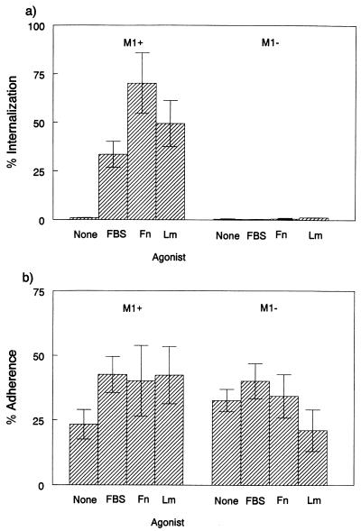
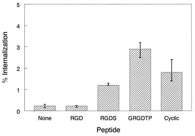
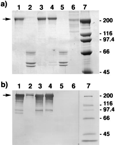
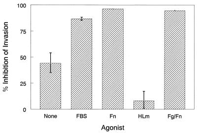
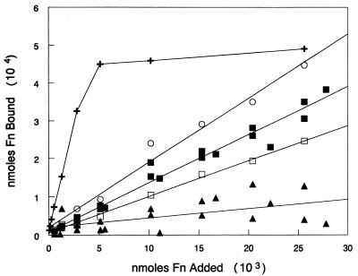
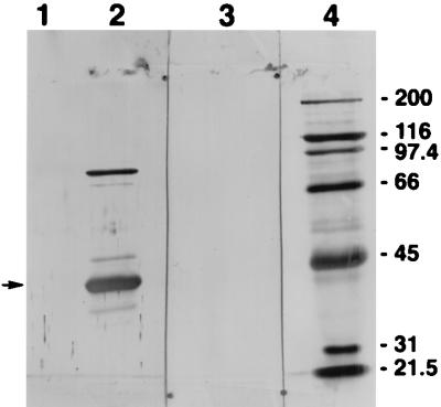
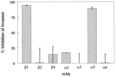
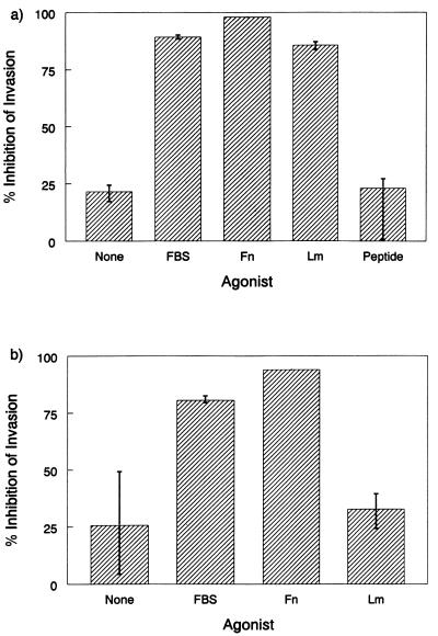
The ability of a serotype M1 strain of Streptococcus pyogenes to efficiently invade A549 human lung epithelial cells was previously shown to be dependent on bacterial exposure to human or bovine serum proteins or synthetic peptides containing the sequence RGD. In this study, stimulation by invasion agonists was determined to be dependent on expression of the streptococcal cell surface protein, M1. Fetal bovine serum (FBS), fibronectin (Fn), the extracellular matrix protein laminin (Lm), and RGD-containing peptides were tested for their abilities to promote epithelial cell invasion and adherence by isogenic M1(+) and M1(-) strains of S. pyogenes. In the absence of an agonist, invasion and adherence were comparable for the two bacterial strains. FBS, Fn, and Lm stimulated invasion of the M1(+) strain as much as 70-fold but failed to significantly affect invasion by the M1(-) mutant. Adherence of the wild-type strain was stimulated by these same agonists. Epithelial cell adherence by the M1(-) strain, however, was unaffected by the presence of Fn or Lm. Several RGD-containing peptides were found to promote invasion independently of M1 expression. Binding of 125I-Fn was reduced 88% by the M1(-) mutation and Fn was found to bind purified M1 protein, suggesting that Fn mediates invasion by direct binding to M1. To determine if host integrins might be involved in internalization of streptococci, several anti-integrin monoclonal antibodies (MAbs) were tested for their abilities to inhibit invasion. Antibody directed against integrin beta1 inhibited FBS-, Fn-, and Lm-mediated invasion but did not abrogate RGD-peptide-stimulated invasion. MAb directed against the epithelial cell Fn receptor, integrin alpha5beta1, inhibited Fn and FBS-mediated invasion but did not specifically inhibit Lm-mediated invasion. These results indicate that S. pyogenes has evolved multiple mechanisms for invasion of eukaryotic cells, at least two of which involve interactions between M1 protein, host integrins, and integrin ligands.


Figures

FIG. 1
FIG. 1
Effects of agonists on adherence and internalization of M1+ and M1 streptococci. M1+ and M1 bacteria were suspended in RPMI 1640 medium (None), or in RPMI 1640 medium containing the indicated agonist, just prior to infection of monolayers. (a) Intracellular invasion. Percent invasion was calculated as (internalized CFU/CFU in the inoculum) × 100. (b) Streptococcal adherence to A549 cells. Percent adherence is expressed as the percentage of total CFU that remained associated with monolayers after three successive washings with buffer. Data are the means ± standard errors of the means from six to nine infected monolayers (two or three experiments, each performed in triplicate).
FIG. 2
FIG. 2
Expression of emm1 is not required for invasion stimulation by RGD peptides. M1 bacteria were suspended in unsupplemented RPMI 1640 medium (None) or in RPMI 1640 medium containing 0.5 mM indicated peptide and then inoculated onto monolayers. Percent invasion was calculated as (internalized CFU/CFU in the inoculum) × 100. Values are mean percentages from a representative experiment in which each assay was performed in triplicate.
FIG. 3
FIG. 3
SDS-PAGE and Western blot analysis of purified invasion agonists. Four micrograms of each of the indicated invasion agonists was electrophoresed through SDS–8% polyacrylamide minigels and then stained with Coomassie blue (a) or transferred to a nitrocellulose membrane and probed with sheep anti-human Fn Ab (b). Lanes: 1, inactive preparation of human Fn; 2, active preparation of human Fg contaminated with Fn; 3, active Fn preparation obtained from Life Technologies; 4, active Fn, purified from the Fg preparation shown in lane 2, by chromatography on gelatin-Sepharose; 5, Fn-depleted Fg (inactive) purified from the lane 2 preparation by gelatin-Sepharose chromatography; 6, HLm (active); 7, molecular weight standards. The numbers to the right indicate the molecular masses of the protein standards in kilodaltons. Fn bands are indicated by the arrows to the left.
FIG. 4
FIG. 4
Invasion inhibition by anti-fibronectin Ab. Sheep anti-human Fn Ab was added to RPMI 1640 medium containing the indicated agonist. M1+ bacteria were added to this medium and to identical media to which no Ab was added. The bacterial suspensions were then added to A549 monolayers. Thereafter, the standard invasion procedure was followed. Percent inhibition was calculated as (CFU recovered from control [no-MAb] wells − CFU recovered from MAb-containing wells/CFU from control wells) × 100. Data are means and ranges from assays performed in triplicate. The Fg, Fn, and HLm preparations used in this experiment were the same as those run in lanes 2, 3, and 6, respectively, of the gels shown in Fig. 3.
FIG. 5
FIG. 5
Schematic representation of the domain structure of M1 protein. (a) Lettered boxes indicate the various domains of M1.0 protein of S. pyogenes AP1 (accession no. 1084197) as previously defined (2). The numbers below the boxes indicate the terminal amino acids residues of the domains. SS, signal sequence; A, N-terminal portion of mature M1 protein comprised of a unique sequence of 91 amino acids; B, B repeat domain, comprised of two repeats of a 28-amino-acid sequence, followed by a 6-amino-acid sequence identical to a portion of the 22-residue repeats, followed by a unique 38-amino-acid sequence; C, C repeat domain comprised of three imperfect repeats of 42, 42, and 31 amino acids; D, D region, C-terminal segment containing the cell wall anchor region, membrane-spanning region, and intracellular portion of M1. Residues 42 to approximately 380 are the extracellular portion of the protein. The amino acid predicted to differ between the M1 proteins of strain 90-226 and S. pyogenes AP1 is indicated at the top. (b) The bar indicates the portion of M1 encoded by plasmid pM42-382. Purified M42-382 is predicted to have a C-terminal glycine residue and an N-terminal formylmethionine not present in native M1 protein.
FIG. 6
FIG. 6
Binding of 125I-labeled Fn by S. pyogenes JRS4 (✚; M6+ protein F+), SAM1 (□; M6+ protein F), AP1 (○; M1+ protein H+), 90-226 (■), and 90-226 emm1::Km (▴). Constant numbers of bacterial cells were suspended in PBSAT, mixed with various amounts of 125I-Fn, and rotated end over end for 2 h at room temperature. Bacterial cells were harvested by centrifugation, and the amount of radioactivity associated with the bacterial pellets was determined. The values plotted are the average counts recovered from duplicate tubes. Nonspecific binding was determined by adding the same amounts of labeled protein to tubes containing no bacteria. Counts recovered from these tubes were subtracted from the values plotted. Data for 90-226 and 90-226 emm1::Km are from three independent sets of experiments, each performed with a different preparation of 125I-labeled Fn.
FIG. 7
FIG. 7
Fibronectin binding to purified M1 protein. Purified M42-382 protein (lanes 2 and 3) and E. coli maltose-binding protein (lane 1) were electrophoresed through SDS–10% acrylamide gels and transferred to a nitrocellulose membrane. The membrane was blocked and then sliced to remove the portion of the membrane containing the lane 3 sample. The portion of the membrane containing the lane 1 and 2 samples was successively incubated with Fn (25 μg/ml) in TBS, sheep anti-human Fn Ab, alkaline phosphatase conjugated to mouse anti-sheep IgG, and finally a chromogenic alkaline phosphatase substrate. The portion of the membrane containing the lane 3 sample was treated similarly except that Fn was omitted from the first membrane incubation. Lane 4 contained molecular weight standards. The numbers to the right indicate molecular masses of the standards in kilodaltons. The arrow indicates the 39.5-kDa M42-382 peptide. The higher-molecular-weight band present in lane 2 appears to be dimerized M42-382 protein.
FIG. 8
FIG. 8
Effects of anti-integrin MAbs on intracellular invasion. MAbs recognizing the indicated integrin subunits were diluted in RPMI 1640 medium containing Fn and incubated with A549 monolayers for 30 min prior to addition of bacteria. Thereafter, the standard assay procedure was followed. Purified MAbs against integrins α5 and β1 were diluted to 0.35 and 0.5 μg/ml, respectively. Other MAbs were diluted 1:50; final concentrations of Abs varied from 4.8 to 38 μg/ml. Percent inhibition was calculated as described in the legend to Fig. 4. Data are means and ranges from assays performed in triplicate.
FIG. 9
FIG. 9
Invasion inhibition by anti-integrin β1 and α5β1 MAbs. MAbs directed against integrin β1 (a) and integrin α5β1 (b) were added to RPMI 1640 medium containing the indicated agonists. M1+ bacteria were added to this medium and to identical media to which MAb was not added. The bacterial suspensions were then inoculated onto A549 monolayers. Thereafter, the standard assay procedure was followed. Percent inhibition was calculated as described in the legend to Fig. 4. Data are mean percentages and ranges from a representative experiment in which each assay was performed in triplicate.

References

    1. Akesson P, Sjoholm A G, Björck L. Protein SIC—a novel extracellular protein of Streptococcus pyogenes interfering with complement function. J Biol Chem. 1996;271:1081–1088. - PubMed
    1. Akesson P, Schmidt K-H, Cooney J, Björck L. M1 protein and protein H: IgGFc- and albumin-binding streptococcal surface proteins encoded by adjacent genes. Biochem J. 1994;300:877–886. - PMC - PubMed
    1. Bayles K W, Wesson C A, Liou L E, Fox L K, Bohach G A, Trumble W R. Intracellular Staphylococcus aureus escapes the endosome and induces apotosis in epithelial cells. Infect Immun. 1998;66:336–342. - PMC - PubMed
    1. Bradford M M. A rapid and sensitive method for the quantitation of microgram quantities of protein utilizing the principle of protein-dye binding. Anal Biochem. 1976;72:248–254. - PubMed
    1. Caixia S, Stewart S, Wayner E, Carter W, Wilkins J. Antibodies to different members of the β1 (CD29) integrins induce homotypic and heterotypic cellular aggregation. Cell Immunol. 1991;138:216–228. - PubMed

Publication types

MeSH terms

LinkOut - more resources