. 1998 Oct;66(10):4602-10.
doi: 10.1128/IAI.66.10.4602-4610.1998.

Surface-associated hsp60 chaperonin of Legionella pneumophila mediates invasion in a HeLa cell model

Affiliations

Surface-associated hsp60 chaperonin of Legionella pneumophila mediates invasion in a HeLa cell model

R A Garduño et al. Infect Immun. 1998 Oct.

Abstract

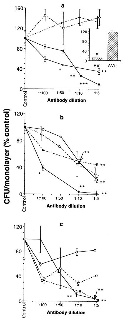
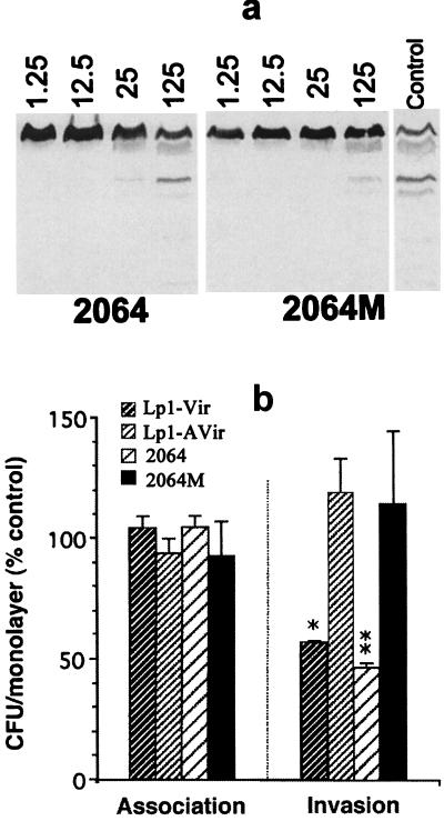
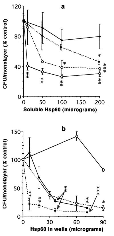
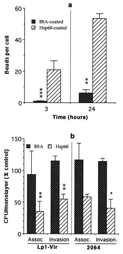
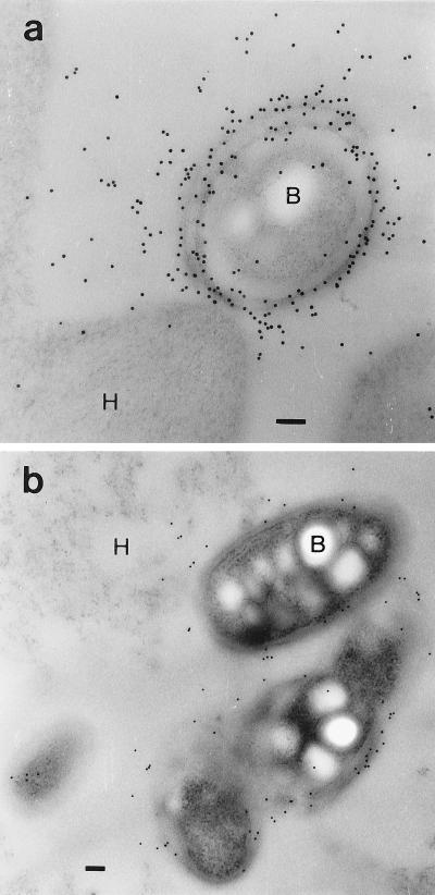
HeLa cells have been previously used to demonstrate that virulent strains of Legionella pneumophila (but not salt-tolerant avirulent strains) efficiently invade nonphagocytic cells. Hsp60, a member of the GroEL family of chaperonins, is displayed on the surface of virulent L. pneumophila (R. A. Garduño et al., J. Bacteriol. 180:505-513, 1988). Because Hsp60 is largely involved in protein-protein interactions, we investigated its role in adherence-invasion in the HeLa cell model. Hsp60-specific antibodies inhibited the adherence and invasiveness of two virulent L. pneumophila strains in a dose-dependent manner but had no effect on the association of their salt-tolerant avirulent derivatives with HeLa cells. A monospecific anti-OmpS (major outer membrane protein) serum inhibited the association of both virulent and avirulent strains of L. pneumophila to HeLa cells, suggesting that while both Hsp60 and OmpS may mediate bacterial association to HeLa cells, only virulent strains selectively displayed Hsp60 on their surfaces. Furthermore, the surface-associated Hsp60 of virulent bacterial cells was susceptible to the action of trypsin, which rendered the bacteria noninvasive. Additionally, pretreatment of HeLa cells with purified Hsp60 or precoating of the plastic surface where HeLa cells attached with Hsp60 reduced the adherence and invasiveness of the two virulent strains. Finally, recombinant Hsp60 covalently bound to latex beads promoted the early association of beads with HeLa cells by a factor of 20 over bovine serum albumin (BSA)-coated beads and competed with virulent strains for association with HeLa cells. Hsp60-coated beads were internalized in large numbers by HeLa cells and remained in tight endosomes that did not fuse with other vesicles, whereas internalized BSA-coated beads, for which endocytic trafficking is well established, resided in more loose or elongated endosomes. Mature intracellular forms of L. pneumophila, which were up to 100-fold more efficient than agar-grown bacteria at associating with HeLa cells, were enriched for Hsp60 on the bacterial surface, as determined by immunolocalization techniques. Collectively, these results establish a role for surface-exposed Hsp60 in invasion of HeLa cells by L. pneumophila.


Figures

FIG. 1
FIG. 1
Effects of antibodies on interactions of different strains of L. pneumophila with HeLa cells. (a) Effects of different dilutions of an Hsp60 rabbit antiserum on the association of the virulent strains (solid lines) Lp1-Vir (○) and 2064 (•), or the avirulent strains (broken lines) Lp1-AVir (○) and 2064M (•), with HeLa cells. The inset indicates the effect of monoclonal antibody GW2X4B8B2H6 on the association of Lp1-Vir and Lp1-AVir with HeLa cells. (b) Effects of different dilutions of an OmpS rabbit antiserum on the association of the four L. pneumophila strains described above. (c) Effects of different dilutions of an Hsp60 antiserum (solid lines) or an OmpS antiserum (broken lines) on the invasiveness of the virulent strain 2064 (•) and the avirulent strain 2064M (○). Graph points represent the means ± standard deviations (vertical bars) from single experiments run in duplicate (n = 2). Asterisks indicate the level of significance of the differences between the indicated graph points and their corresponding controls at P values of ≤0.05 (∗), ≤0.01 (∗∗), and ≤0.001 (∗∗∗). P values were calculated by comparing mean CFU/monolayer values (not the relative percentages), and their corresponding standard deviations, in Student t tests.
FIG. 2
FIG. 2
Effects of trypsin treatment on interactions of different strains of L. pneumophila with HeLa cells. (a) Immunoblot showing differential proteolysis of Hsp60 in whole cells of virulent 2064 and avirulent 2064M strains. Numbers above the lanes indicate the amounts (in micrograms) of trypsin added to ∼109 whole bacterial cells (in a final volume of 100 μl) to effect the proteolysis of exposed proteins. Coomassie blue staining detected no apparent degradation of other proteins. The lane marked Control shows the degradation of Hsp60 in lysates of 2064M to which no trypsin inhibitor was added. (b) Levels of association and invasion of L. pneumophila strains treated with trypsin before infection of HeLa cells. For levels of significance, see the legend to Fig. 1.
FIG. 3
FIG. 3
Effects of purified Hsp60 on interactions of our virulent strains of L. pneumophila with HeLa cells. (a) Competition for Hsp60 binding sites by soluble Hsp60 added to the HeLa cell supernatant before infection with the virulent strain Lp1-Vir (○) or 2064 (•). Cell association (solid lines) and invasion (broken lines) were determined in standard invasion assays. (b) Receptor modulation assays carried in the presence of different amounts of substratum-bound Hsp60. Symbols are as described for panel a. Points represent means ± standard deviations (vertical bars) of single experiments run in duplicate. Statistical significance was calculated in relation to controls run in the presence of soluble BSA (a) or substratum-bound BSA (b). Levels of significance are indicated as detailed in the legend to Fig. 1.
FIG. 4
FIG. 4
Interaction of HeLa cells with latex beads coated with Hsp60 or BSA. (a) Number of coated beads/cell in HeLa monolayers to which beads were added at a bead-to-HeLa cell ratio of 100:1 to 200:1. (b) Competition for available binding sites between Hsp60-coated beads (Hsp60) or BSA-coated beads (BSA) and our virulent L. pneumophila strains. Columns represent means + standard deviations (vertical bars) of single experiments run in triplicate (a) or in duplicate (b). In panel b, the percentages were calculated in relation to a common control to which no beads were added. Levels of significance are as detailed in the legend to Fig. 1 and refer to differences between the corresponding BSA and Hsp60 columns.
FIG. 5
FIG. 5
Ultrastructural view of the interaction of coated beads with HeLa cells. Electron micrographs show intracellular Hsp60-coated beads (a to c) or BSA-coated beads (d) taken up by HeLa cells. Micrographs were taken from specimens fixed 3 h (a and b) or 24 h (c and d) after addition of the beads. Arrowheads in panel b point to the tight association between the endosomal membrane and the surface of beads. Arrows in panel d point to the loose endosomes containing BSA-coated beads. V, unidentified vesicle commonly found in association with endosomes containing Hsp60-coated beads. Bars represent 1 μm in all panels.
FIG. 6
FIG. 6
Immunolocalization of Hsp60 or OmpS epitopes in mature forms of L. pneumophila 2064 infecting HeLa cells. Ultrathin sections of HeLa cells infected with mature bacteria were immunolabeled with rabbit anti-Hsp60 serum (a) or rabbit anti-OmpS serum (b) and a secondary antibody (goat anti-rabbit immunoglobulin G) conjugated to 10-nm gold particles (Sigma Immunochemicals). Bars represent 0.1 μm in both panels. H, HeLa cells; B, mature bacteria isolated from infected HeLa cells.

References

    1. Abu Kwaik Y. The phagosome containing Legionella pneumophila within the protozoan Hartmanella vermiformis is surrounded by the rough endoplasmic reticulum. Appl Environ Microbiol. 1996;62:2022–2028. - PMC - PubMed
    1. Abu Kwaik Y, Fields B S, Engleberg C. Protein expression by the protozoan Hartmanella vermiformis upon contact with its bacterial parasite Legionella pneumophila. Infect Immun. 1994;62:1860–1866. - PMC - PubMed
    1. Andrews H L, Vogel J P, Isberg R R. Identification of linked Legionella pneumophila genes essential for intracellular growth and evasion of the endocytic pathway. Infect Immun. 1998;66:950–958. - PMC - PubMed
    1. Berger K H, Merriam J J, Isberg R R. Altered intracellular targeting properties associated with mutations in the Legionella pneumophila dotA gene. Mol Microbiol. 1994;14:809–822. - PubMed
    1. Bozue J A, Johnson W. Interaction of Legionella pneumophila with Acanthamoeba castellanii: uptake by coiling phagocytosis and inhibition of phagosome-lysosome fusion. Infect Immun. 1996;64:668–673. - PMC - PubMed

Publication types

MeSH terms

LinkOut - more resources