. 1998 Oct;66(10):4669-75.
doi: 10.1128/IAI.66.10.4669-4675.1998.

Induction of adrenomedullin mRNA and protein by lipopolysaccharide and paclitaxel (Taxol) in murine macrophages

Affiliations

Induction of adrenomedullin mRNA and protein by lipopolysaccharide and paclitaxel (Taxol) in murine macrophages

M Zaks-Zilberman et al. Infect Immun. 1998 Oct.

Abstract

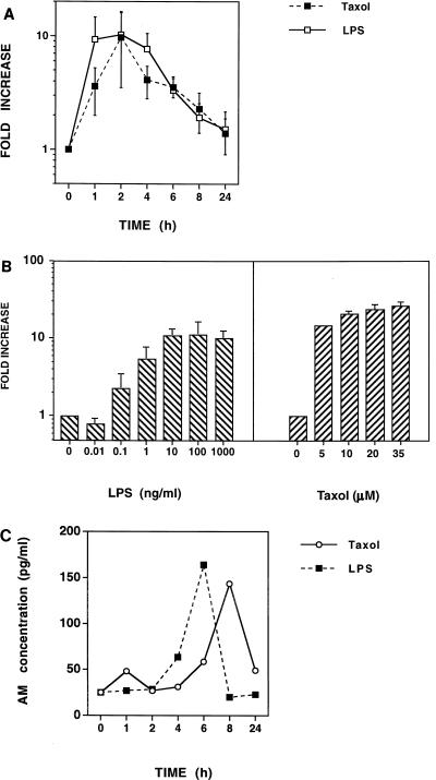
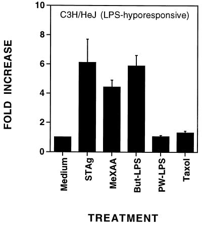
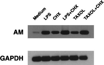
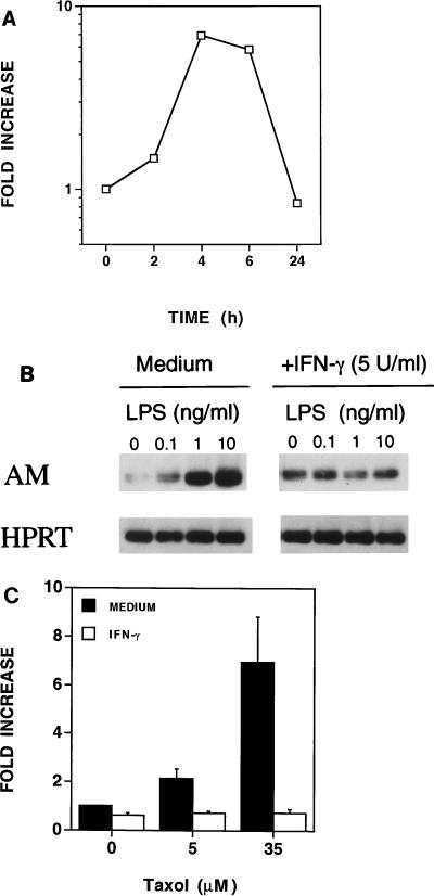
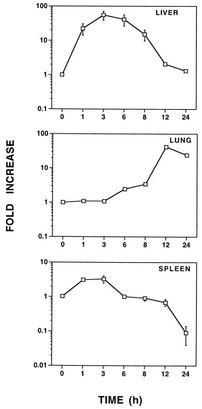
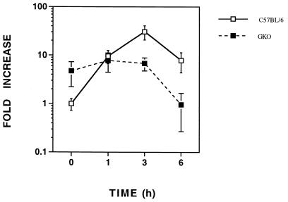
Lipopolysaccharide (LPS), a potent inflammatory stimulus derived from the outer membrane of gram-negative bacteria, has been implicated in septic shock. Plasma levels of adrenomedullin (AM), a potent vasorelaxant, are increased in septic shock and possibly contribute to the characteristic hypotension. As macrophages play a central role in the host response to LPS, we studied AM production by LPS-stimulated macrophages. When peritoneal exudate macrophages from C3H/OuJ mice were treated with protein-free LPS (100 ng/ml) or the LPS mimetic paclitaxel (Taxol; 35 microM), an approximately 10-fold increase in steady-state AM mRNA levels was observed, which peaked between 2 and 4 h. A three- to fourfold maximum increase in the levels of immunoreactive AM protein was detected after 6 to 8 h of stimulation. While LPS-hyporesponsive C3H/HeJ macrophages failed to respond to protein-free LPS with an increase in steady-state AM mRNA levels, increased levels were observed after stimulation of these cells with a protein-rich (butanol-extracted) LPS preparation. In addition, increased AM mRNA was observed following treatment of either C3H/OuJ or C3H/HeJ macrophages with soluble Toxoplasma gondii tachyzoite antigen or the synthetic flavone analog 5, 6-dimethylxanthenone-4-acetic acid. Gamma interferon also stimulated C3H/OuJ macrophages to express increased AM mRNA levels yet was inhibitory in the presence of LPS or paclitaxel. In vivo, mice challenged intraperitoneally with 25 microg of LPS exhibited increased AM mRNA levels in the lungs, liver, and spleen; the greatest increase (>50-fold) was observed in the liver and lungs. Thus, AM is produced, by murine macrophages, and furthermore, LPS induces AM mRNA in vivo in a number of tissues. These data support a possible role for AM in the pathophysiology of sepsis and septic shock.


Figures

FIG. 1
FIG. 1
PW-LPS and paclitaxel induce AM mRNA and protein synthesis in C3H/OuJ macrophages. (A) Kinetics of PW-LPS- and paclitaxel-induced AM mRNA expression. C3H/OuJ macrophages were cultured for the indicated times with medium, 100-ng/ml PW-LPS, or 35 μM paclitaxel. mRNA was isolated, and AM and HPRT mRNAs were detected by RT-PCR. The data represent the arithmetic mean ± the standard error of the mean (seven separate experiments). (B) Dose-dependent induction of AM mRNA. C3H/OuJ macrophages were cultured for 2 h with medium or with the indicated concentrations of PW-LPS or paclitaxel. mRNA was isolated, and AM and HPRT mRNAs were detected by RT-PCR. The data represent the arithmetic mean ± the standard error of the mean (four separate experiments). (C) Kinetics of PW-LPS- and paclitaxel-induced AM secretion. C3H/OuJ macrophages were cultured for the indicated times with medium, 100-ng/ml LPS, or 35 μM paclitaxel. Macrophage culture supernatants were analyzed by RIA for the presence of immunoreactive AM. Data were derived from a representative of three experiments. When not visible, bars indicating the standard error of the mean are smaller than the symbol.
FIG. 2
FIG. 2
Neither PW-LPS nor paclitaxel induced AM gene expression in LPS-hyporesponsive C3H/HeJ macrophages in vitro. C3H/HeJ macrophages were treated for 4 h with medium, 5-μg/ml STAg, 10-μg/ml MeXAA, 5-μg/ml But-LPS, 100-ng/ml PW-LPS, or 35 μM paclitaxel. The data represent the arithmetic mean ± the standard error of the mean of four experiments. When not visible, bars indicating the standard error of the mean are smaller than the symbol.
FIG. 3
FIG. 3
De novo protein synthesis is not required for PW-LPS- or paclitaxel-induced AM mRNA production. C3H/OuJ macrophages were treated for 2 h with either medium, 100-ng/ml PW-LPS, 5-μg/ml CHX, both 100-ng/ml PW-LPS and 5-μg/ml CHX, 35 μM paclitaxel, or both 35 μM paclitaxel and 5-μg/ml CHX. RNA was isolated, and AM and GAPDH mRNAs were detected by RT-PCR. A representative of three Southern blots is shown.
FIG. 4
FIG. 4
Regulation of AM mRNA expression by IFN-γ in C3H/OuJ macrophages. (A) IFN-γ upregulates AM mRNA expression. C3H/OuJ macrophages were cultured for the indicated times with medium or 5-U/ml IFN-γ. These data were derived from a representative of two experiments. (B) IFN-γ down-regulates PW-LPS-induced AM mRNA expression. C3H/OuJ macrophages were cultured for 4 h in the presence of medium only or increasing concentrations of PW-LPS in the absence or presence of 5-U/ml IFN-γ. A representative of three Southern blots is shown. (C) IFN-γ down-regulates paclitaxel-induced AM mRNA expression. C3H/OuJ macrophages were cultured for 4 h in the presence of medium only, 5 μM paclitaxel, or 35 μM paclitaxel, in the absence or presence of 5-U/ml IFN-γ. These data represent the arithmetic mean ± the standard error of the mean of three separate experiments.
FIG. 5
FIG. 5
LPS augments AM mRNA expression in vivo. C57BL/6 mice were injected i.p. with 25 μg of LPS. These data are the mean fold increase in AM mRNA expression ± the standard error of the mean from four to eight individual mice at each time point. Means are expressed relative to that of the saline-injected control (t = 0), which was arbitrarily assigned a value of 1. When not visible, bars indicating the standard error of the mean are smaller than the symbol.
FIG. 6
FIG. 6
Endogenous IFN-γ regulates LPS-induced AM mRNA in vivo. GKO and C57BL/6 mice were injected i.p. with 25 μg of LPS, and LPS-induced AM mRNA was quantified in the liver. These data are the arithmetic mean ± the standard error of the mean for five to eight mice at each time point.

Similar articles

Cited by

References

    1. Abraham E, Wunderink R, Silverman H, Perl T M, Nasraway S, Levy H, Bone R, Wenzel R P, Balk R, Allred R. Efficacy and safety of monoclonal antibody to human tumor necrosis factor alpha in patients with sepsis syndrome. A randomized, controlled, double-blind, multicenter clinical trial. TNF-alpha MAb Sepsis Study Group. JAMA. 1995;273:934–941. - PubMed
    1. Atwell G J, Rewcastle G W, Baguley B C, Denny W A. Potential antitumor agents. 60. Relationships between structure and in vivo colon 38 activity for 5-substituted 9-oxoxanthene-4-acetic acids. J Med Chem. 1990;33:1375–1379. - PubMed
    1. Barber S A, Fultz M J, Salkowski C A, Vogel S N. Differential expression of interferon regulatory factor 1 (IRF-1), IRF-2, and interferon consensus sequence binding protein genes in lipopolysaccharide (LPS)-responsive and LPS-hyporesponsive macrophages. Infect Immun. 1995;63:601–608. - PMC - PubMed
    1. Bogdan C, Ding A. Taxol, a microtubule-stabilizing antineoplastic agent, induces expression of tumor necrosis factor alpha and interleukin-1 in macrophages. J Leukocyte Biol. 1992;52:119–121. - PubMed
    1. Bone R C. A critical evaluation of new agents for the treatment of sepsis. JAMA. 1991;266:1686–1691. - PubMed

Publication types