. 1998 Oct;66(10):4676-89.
doi: 10.1128/IAI.66.10.4676-4689.1998.

Filament tip-associated antigens involved in adherence to and invasion of murine pulmonary epithelial cells in vivo and HeLa cells in vitro by Nocardia asteroides

Affiliations

Filament tip-associated antigens involved in adherence to and invasion of murine pulmonary epithelial cells in vivo and HeLa cells in vitro by Nocardia asteroides

B L Beaman et al. Infect Immun. 1998 Oct.

Abstract

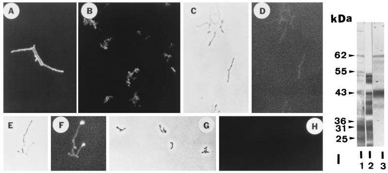
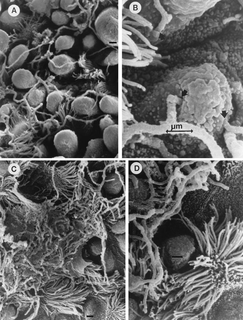
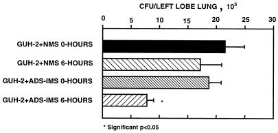
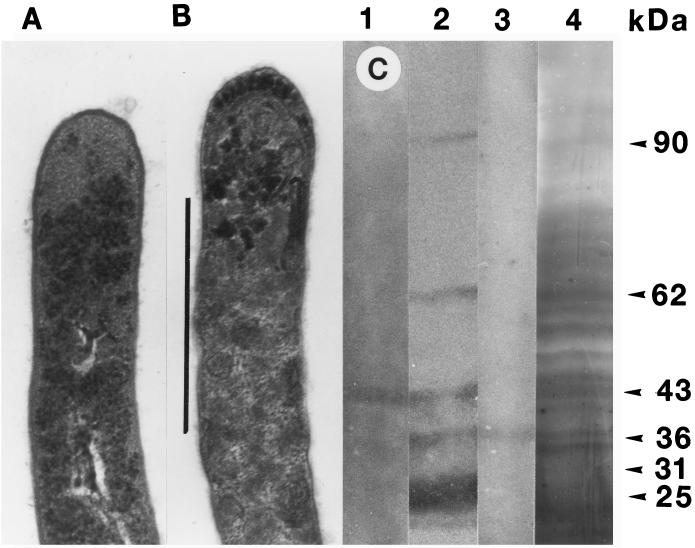
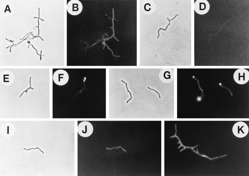
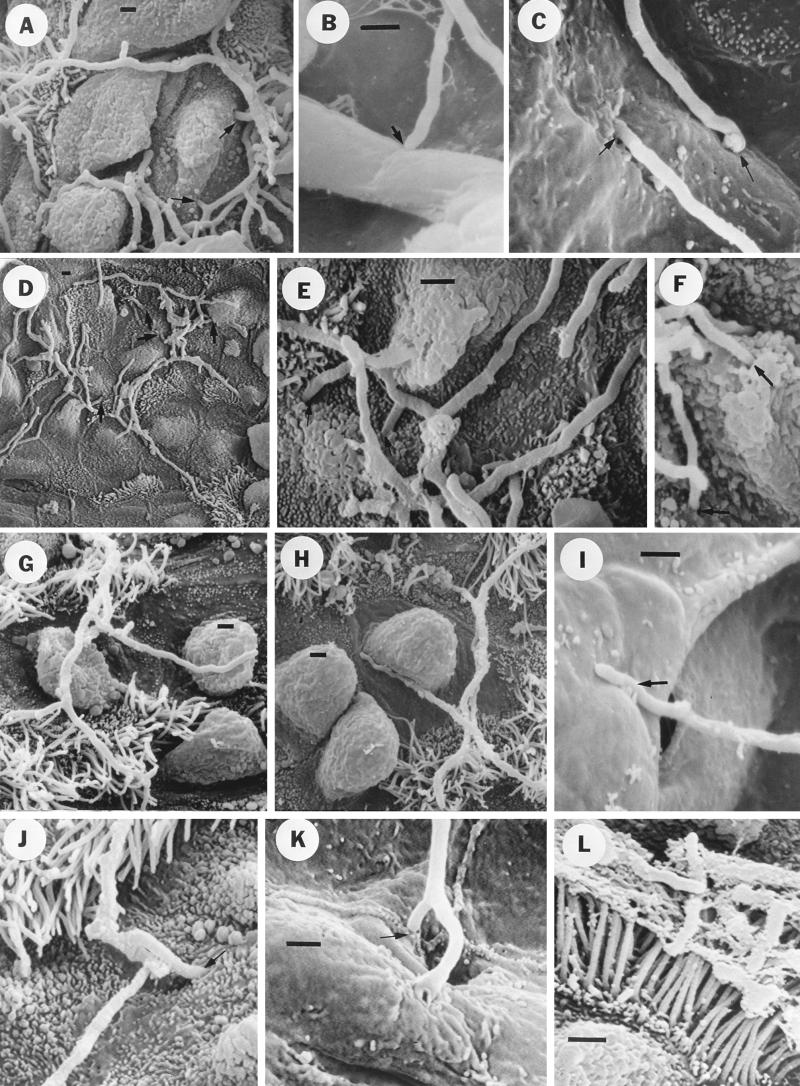
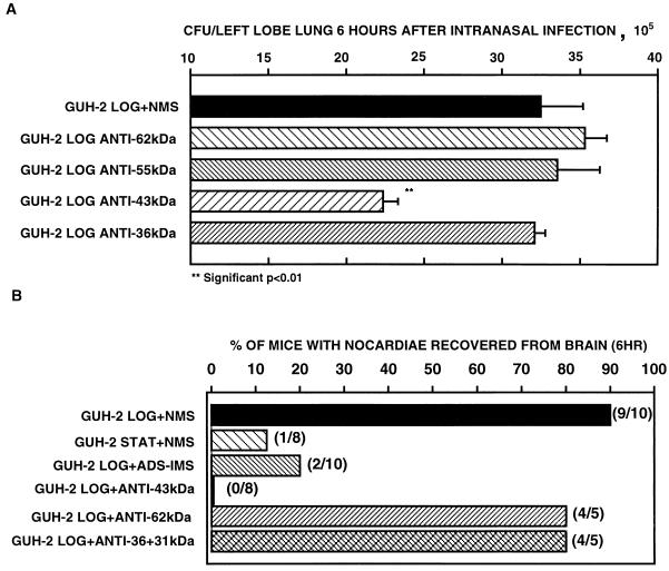
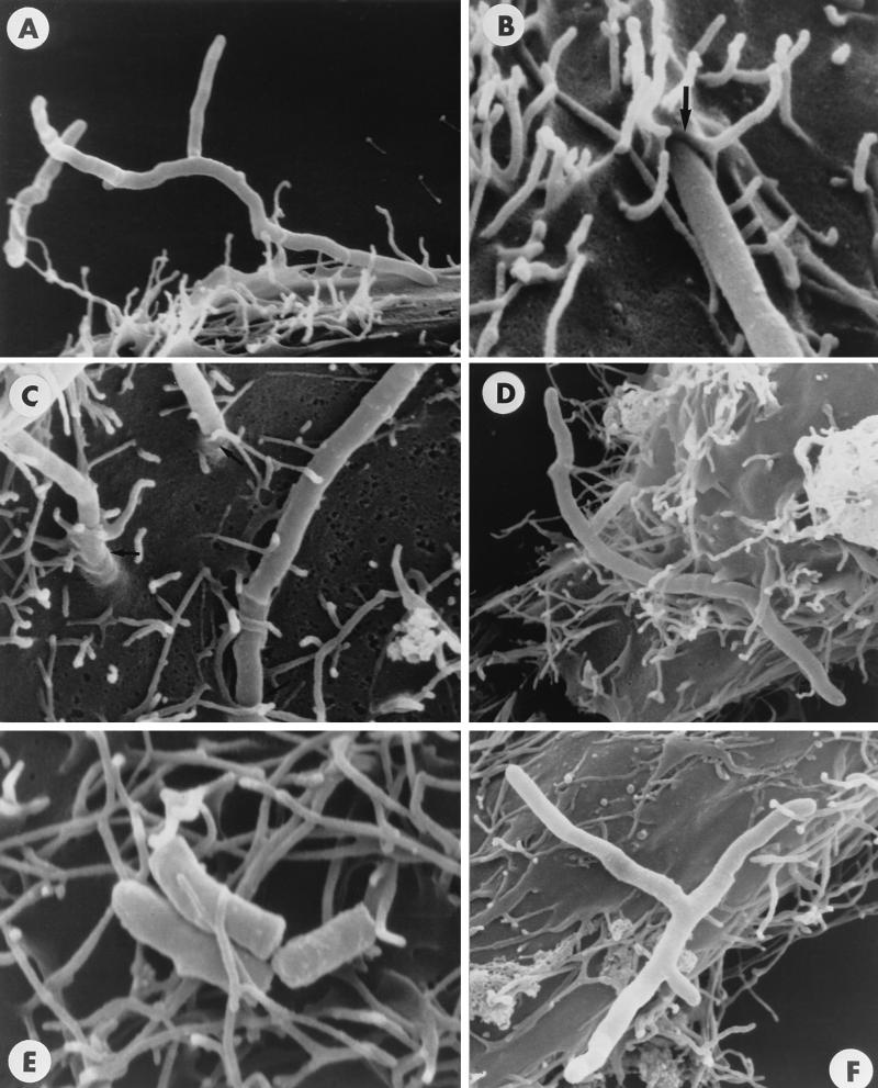
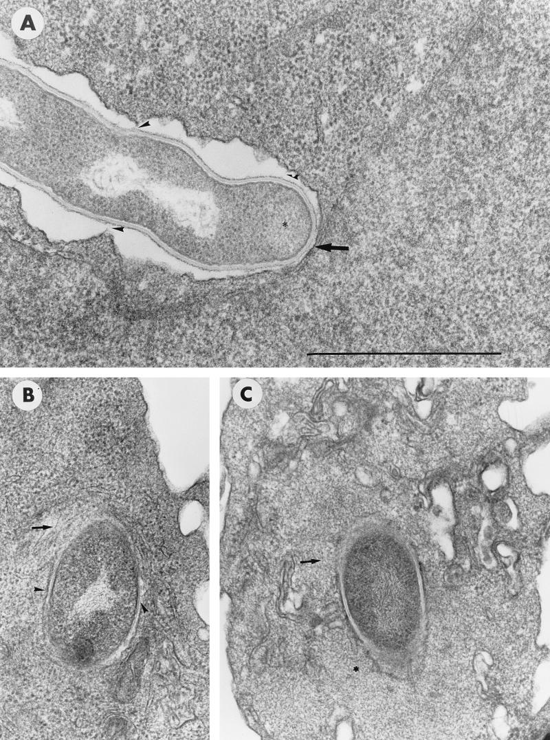
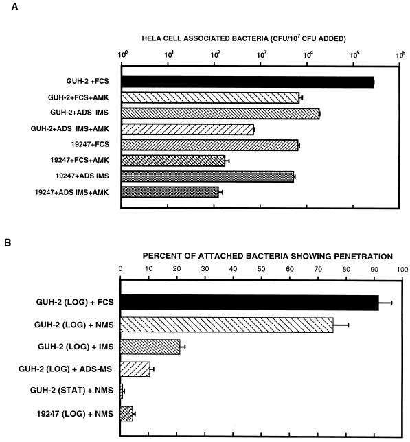
The interactions of Nocardia asteroides GUH-2 with pulmonary epithelial cells of C57BL/6 mice and with HeLa cells were studied. Electron microscopy demonstrated that only the tips of log-phase cells penetrated pulmonary epithelial cells following intranasal administration, and nocardiae were recovered from the brain. Coccobacillary cells neither invaded nor disseminated. Serum from immunized mice (IMS) decreased attachment to and penetration of pulmonary epithelial cell surfaces by log-phase GUH-2 and inhibited spread to the brain. IMS was adsorbed against stationary-phase cells. Western immunoblots suggested that this adsorbed IMS was reactive primarily with 43- and 62-kDa proteins. Immunofluorescence showed that adsorbed IMS preferentially labeled the tips of log-phase GUH-2 cells. Since this IMS was reactive to culture filtrate antigens, several of these proteins were cut from gels, and mice were immunized. Sera against 62-, 55-, 43-, 36-, 31-, and 25-kDa antigens were obtained. The antisera against the 43- and 36-kDa proteins labeled the filament tips of GUH-2 cells. Only the antiserum against the 43-kDa antigen increased pulmonary clearance, inhibited apical attachment to and penetration of pulmonary epithelial cells, and prevented spread to the brain. An in vitro model with HeLa cells demonstrated that the tips of log-phase cells of GUH-2 adhered to and penetrated the surface of HeLa cells. Invasion assays with amikacin treatment demonstrated that nocardiae were internalized. Adsorbed IMS blocked attachment to and invasion of these cells. These data suggested that a filament tip-associated 43-kDa protein was involved in attachment to and invasion of pulmonary epithelial cells and HeLa cells by N. asteroides GUH-2.


Figures

FIG. 1
FIG. 1
Indirect immunofluorescence microscopy of N. asteroides GUH-2 treated with various murine antisera and fluorescein isothiocyanate-conjugated goat antimouse immunoglobulin. Fluorescence was visualized by using epifluorescence illumination on a Ziess research microscope. (A) Log-phase (16-h) GUH-2 incubated with IMS. (B) Stationary-phase (120-h) GUH-2 incubated with IMS. (C) Phase-contrast micrograph of log-phase GUH-2 treated with NMS. (D) Immunofluorescence of the same bacteria shown in panel C. There was no specific reactivity (negative control), but slight autofluorescence was evident. (E) Phase-contrast micrograph of log-phase GUH-2 treated with ADS-IMS. (F) Immunofluorescence of the same bacterium shown in panel E. Note strong specific fluorescence localized at the filament tips. (G) Phase-contrast micrograph of stationary-phase GUH-2 treated with ADS-IMS. (H) Immunofluorescence of the same bacteria shown in panel G. Note the total absence of fluorescence. (I) Western immunoblot against SDS-soluble proteins of log-phase cells of N. asteroides GUH-2. Lane 1, culture filtrate antigens incubated with IMS; lane 2, SDS-solubilized cell walls incubated with IMS; lane 3, SDS-extracted proteins from live, log-phase GUH-2 incubated with ADS-IMS. Molecular mass markers are located relative to the migration of specific protein standards of known molecular mass.
FIG. 2
FIG. 2
Scanning electron micrographs showing the effect of ADS-IMS on the invasion of nonciliated bronchiolar epithelial cells by log-phase cells of N. asteroides GUH-2 in C57BL/6 mice. Bars, 1 μm. (A) Low-magnification view showing nocardial filaments among the Clara cells and not specifically associated with ciliated cells (compare to panel C). Treatment was with NMS. (B) High-magnification view of an region adjacent to that in panel A, showing nocardial filaments penetrating into epithelial cells (arrows). Treatment was with NMS. (C) Low-magnification view showing nocardial filaments highly associated with ciliated cells on the bronchiolar surface (treatment was with ADS-IMS). Note that most of the filaments appear to be embedded in a material that may be mucus lying on the surface, whereas masses of nocardial filaments embedded in this mucous material were not observed in the lungs of NMS-treated controls (compare panels A and C). (D) High-magnification view showing nocardial filaments on the bronchiolar surface and associated with ciliated cells. Note that freely associated filaments appear to be only longitudinally adherent to the surface, and filament tips did not appear to be either adhering to or penetrating epithelial cells. Treatment was with ADS-IMS.
FIG. 3
FIG. 3
Pulmonary clearance of log-phase cells of N. asteroides GUH-2 following incubation with ADS-IMS, showing the effect of ADS-IMS 6 h after i.n. administration to C57BL/6 mice. Error bars represent standard errors (n = 5).
FIG. 4
FIG. 4
Effect of SDS extraction on log-phase cells of N. asteroides GUH-2. (A) Electron microscopy of the tip of a log-phase GUH-2 cell grown in BHI broth without extraction. (B) Electron microscopy of the tip of a log-phase GUH-2 cell grown in BHI broth boiled in 1% SDS for 1 h. Bar, 1 μm. The cells in panels A and B are at the same magnification. (C) Western blot characterization of the SDS-extracted components. Lane 1, Western blot against the 43-kDa protein antigen (anti-43-kDa murine serum); lane 2, Western blot with IMS on the SDS extract; lane 3, Western blot against the 36-kDa protein antigen (anti-36-kDa murine serum); lane 4, silver stain after PAGE of the SDS extract from boiling log-phase GUH-2.
FIG. 5
FIG. 5
Indirect immunofluorescence microscopy of N. asteroides GUH-2 treated with various murine antisera and fluorescein isothiocyanate-conjugated goat anti-mouse immunoglobulin. Fluorescence was visualized with epifluorescence illumination on a Ziess research microscope. (A) Phase-contrast micrograph of log-phase GUH-2 treated with anti-62-kDa antiserum. (B) Immunofluorescence of the same bacteria shown in panel A. Note the specific fluorescence localized along only portions of nocardial filaments, with no enhanced apical immunoreactivity. (C) Phase-contrast micrograph of log-phase GUH-2 treated with anti-55-kDa antiserum. (D) Immunofluorescence of the same bacteria shown in panel C. Only slight autofluorescence was evident. (E) Phase-contrast micrograph of log-phase GUH-2 treated with anti-43-kDa antiserum. (F) Immunofluorescence of the same bacterium shown in panel E. Note the strong specific fluorescence localized at the filament tip. (G) Phase-contrast micrograph of log-phase GUH-2 treated with anti-36-kDa antiserum. (H) Immunofluorescence of the same bacteria shown in panel G. Note the strong specific fluorescence localized at the filament tips. (I) Phase-contrast micrograph of log-phase GUH-2 treated with anti-31-kDa antiserum. (J) Immunofluorescence of the same bacterium shown in panel I. Note the specific fluorescence along the nocardial filament, with no enhanced apical immunoreactivity. (K) Immunofluorescence of log-phase GUH-2 treated with anti-25-kDa antiserum. (This 25-kDa protein appears to be nocardial superoxide dismutase [13].) Specific fluorescence was shown along the nocardial filament, with no enhanced apical immunoreactivity.
FIG. 6
FIG. 6
Scanning electron micrographs showing the effects of antisera against selected surface-associated protein antigens of N. asteroides GUH-2 on invasion of bronchiolar and alveolar epithelial cells. Bars, 1 μm. (A) Nocardial filaments incubated with NMS (control) among bronchiolar Clara cells, showing nocardial penetration into these epithelial cells (arrows). (B) Nocardial filament incubated with NMS (control), showing apical adherence to an alveolar septum (arrow). (C) Nocardial filaments incubated with NMS (control), showing both apical adherence to and penetration of the alveolar surface (arrows). (D) Low-magnification view showing nocardial filaments incubated with anti-62-kDa antiserum among the Clara cells and not specifically associated with ciliated cells (compare with Fig. 2A). (E) High-magnification view showing nocardial filaments (treated with anti-62-kDa antiserum) penetrating into epithelial cells (arrows) (compare to controls). (F) Nocardial filaments incubated with anti-55-kDa antiserum penetrating into bronchiolar epithelial cells (arrows), as observed for NMS controls (panel A). (G) Low-magnification view showing nocardial filaments incubated with anti-43-kDa antiserum among bronchiolar cells. Note the apparent association with cilia and possible longitudinal adherence to a Clara cell (compare to panel D). (H) Same as panel G, but a different region showing tighter longitudinal adherence of nocardial filament to a Clara cell. Note the absence of apical adherence to and penetration of bronchiolar epithelial cells. (I) Nocardial filament incubated with anti-43-kDa antiserum, exhibiting longitudinal adherence to an alveolar cell (arrow) (compare to panel B). (J) Nocardial filament incubated with anti-36-kDa antiserum penetrating into a bronchiolar epithelial cell (arrow), as observed for the NMS control (panel A). (K) A branched nocardial filament incubated with anti-36-kDa antiserum, showing apical penetration of the alveolar surface (arrow). (L) Stationary-phase cells of GUH-2 appear to be embedded in a mucous material lying on the surface of ciliated bronchiolar epithelial cells. Note that apical adherence to and penetration of pulmonary epithelial cells were not observed.
FIG. 7
FIG. 7
(A) Effects of selected antisera against individual secreted protein antigens of N. asteroides GUH-2 on pulmonary clearance 6 h after i.n. administration. (B) Effects of different antisera on the spread of N. asteroides GUH-2 to the brains of C57BL/6 mice 6 h after i.n. administration of approximately 2 × 10⁶ CFU/left lobe of lung (positive mice/total mice studied). LOG, log phase; STAT, stationary phase.
FIG. 8
FIG. 8
Scanning electron micrographs showing surface interactions of N. asteroides GUH-2 incubated with HeLa cells for 1 h. (A) Apical attachment of log-phase GUH-2 to the surface of a HeLa cell (incubated with FCS and NMS). (B) The arrow shows apical penetration of the HeLa cell surface by a log-phase filament of GUH-2 (incubated with FCS only). (C) Tips of three log-phase filaments of GUH-2 penetrating the surface of the HeLa cell (incubated with FCS and NMS). (D) Longitudinal attachment of a log-phase filament of GUH-2 (incubated with FCS and NMS). (E) Longitudinal attachment of stationary-phase GUH-2 to the surface of HeLa cells (incubated with FCS and NMS). Neither apical adherence nor penetration of the HeLa cell surface was observed. (F) Log-phase filament of GUH-2 incubated with FCS and ADS-IMS prior to incubation with HeLa cells. Few bacteria showed apical penetration. Magnifications are indicated by the nocardial filament diameter of approximately 0.5 μm.
FIG. 9
FIG. 9
Transmission electron microscopy of log-phase cells of N. asteroides GUH-2 in FCS incubated for 1 h with HeLa cells. (A) Filament tip penetrating the surface of the HeLa cell as in Fig. 1B. The arrow indicates a tight association of electron-dense material on the filament apex with the cytoplasmic membrane of the HeLa cell. Arrowheads indicate apparent sites of attachment between the outer filament wall and the HeLa cell membrane. Bar, 1.0 μm. (B) Nocardial filament localized within the HeLa cell. The arrow indicates microfilaments surrounding the internalized nocardia, and the arrowhead indicates a tight association of the host cell membrane surrounding the nocardial cell. (C) A different preparation showing the same features of internalized log-phase cells of GUH-2 in HeLa cells as in panel B. The asterisks indicate the presence of a granular material. In panel A, the asterisk indicates activity seen with actively growing filament tip. In panel C, the asterisk indicates the host cell, showing a region of increased metabolic activity.
FIG. 10
FIG. 10
(A) Quantitation of the relative numbers (log10) of log-phase cells of an invasive strain (GUH-2) and a noninvasive strain (ATCC 19247) of N. asteroides adhering to and invading HeLa cells as determined with an amikacin (AMK) killing assay. Error bars indicate standard errors. (B) Quantitative scanning electron microscopy estimation of the effects of different murine sera on apical attachment to and penetration of HeLa cells by N. asteroides. The average percentage of attached nocardiae showing evidence of penetration of the HeLa cell surface (calculated as total number of tips showing penetration/total number of bacteria attached) is shown. Error bars indicate standard errors (three separate experiments). LOG, log phase; STAT, stationary phase (see Materials and Methods).

References

    1. Arruda S, Bomfim G, Knights R, Huima-Byron T, Riley L W. Cloning of an M. tuberculosis DNA fragment associated with entry and survival inside cells. Science. 1993;261:1454–1456. - PubMed
    1. Baba T, Nishiuchi Y, Yano I. Composition of mycolic acid molecular species as a criterion in nocardial classification. Int J Syst Bacteriol. 1997;47:795–801.
    1. Badger J L, Miller V L. Expression of invasion and motility are coordinately regulated in Yersinia enterocolitica. J Bacteriol. 1998;180:793–800. - PMC - PubMed
    1. Beaman B L. Differential binding of Nocardia asteroides in the murine lung and brain suggests multiple ligands on the nocardial surface. Infect Immun. 1996;64:4859–4862. - PMC - PubMed
    1. Beaman B L. Interaction of Nocardia asteroides at different phases of growth with in vitro-maintained macrophages obtained from the lungs of normal and immunized rabbits. Infect Immun. 1979;26:355–361. - PMC - PubMed

Publication types

Substances

LinkOut - more resources