. 1998 Oct;66(10):4700-10.
doi: 10.1128/IAI.66.10.4700-4710.1998.

Identification of two Shigella flexneri chromosomal loci involved in intercellular spreading

Affiliations

Identification of two Shigella flexneri chromosomal loci involved in intercellular spreading

M Hong et al. Infect Immun. 1998 Oct.

Abstract

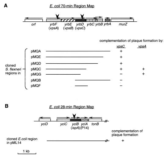
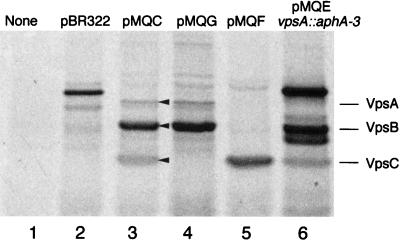
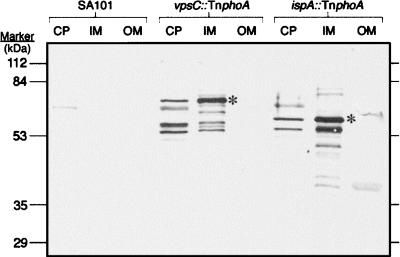
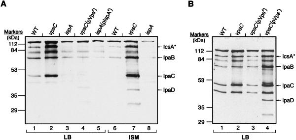
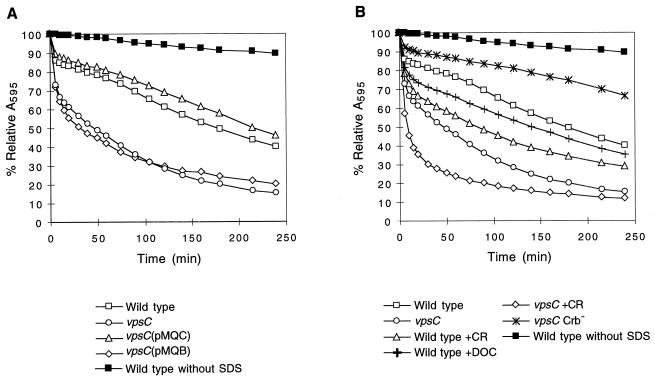
The ability of Shigella flexneri to multiply within colonic epithelial cells and spread to adjacent cells is essential for production of dysentery. Two S. flexneri chromosomal loci that are required for these processes were identified by screening a pool of TnphoA insertion mutants. These mutants were able to invade cultured epithelial cells but could not form wild-type plaques. Analysis of the nucleotide sequence indicated that the sites of TnphoA insertion were within two different regions that are almost identical to Escherichia coli K-12 chromosomal sequences of unknown functions. One region is located at 70 min on the E. coli chromosome, upstream of murZ, while the other is at 28 min, downstream of tonB. The mutant with the insertion at 70 min was named vpsC because it showed an altered pattern of virulence protein secretion. The vpsC mutant formed pinpoint-sized plaques, was defective in recovery from infected tissue culture cells, and was sensitive to lysis by the detergent sodium dodecyl sulfate. Recombinant plasmids carrying the S. flexneri vpsA, -B, and -C genes complemented all of the phenotypes of the vpsC mutant. A mutation in vpsA resulted in the same phenotype as the vpsC mutation, suggesting that these two genes are part of a virulence operon in S. flexneri. The mutant with the insertion at 28 min was interrupted in the same open reading frame as S. flexneri ispA. This ispA mutant could not form plaques and was defective in bacterial septation inside tissue culture cells.


Figures

FIG. 1
FIG. 1
Maps of the 70- and 28-min regions of E. coli and S. flexneri. These maps are in accordance with E. coli chromosomal sequences at the 70-min (A) and 28-min (B) regions to which S. flexneri sequences are highly homologous (; also this study) (GenBank accession no. U18997). Boxes with different patterns represent ORFs or genes that were characterized. Horizontal arrows above ORFs indicate the putative directions of transcription. yrb and yci designations are those of Plunkett (47) and Stoltzfus et al. (59). vps genes were named in this study, and ispA was identified by Mac Síomóin et al. (27), while P14 was named by Postle and Good (50). SA2122 had the TnphoA inserted after the codon for isoleucine at amino acid position 127 of VpsC, VpsA is interrupted in SA4100 by insertion of aphA-3 after the codon for arginine at amino acid position 241, and SA2054 had TnphoA inserted after the codon for threonine at amino acid position 65 of IspA. Insertions are indicated by large arrowheads. Lines beneath the maps represent the wild-type regions cloned in the indicated recombinant plasmids. All pMQ plasmids carried wild-type S. flexneri sequences, while pML14 had the wild-type E. coli sequence. The complementation results in the plaque assays are shown.
FIG. 2
FIG. 2
In vitro translation of proteins encoded by plasmids from the vpsABC region. [35S]methionine-labeled proteins produced in an E. coli extract were separated on an SDS–12.5% polyacrylamide gel and visualized by autoradiography. Plasmid DNA templates were as follows: none (lane 1), pBR322 (lane 2), pMQC (vpsABC) (lane 3), pMQG (vpsAB) (lane 4), pMQF (vpsC) (lane 5), pMQEvpsA::aphA-3 (vpsBC) (lane 6). The arrowheads show the positions of the VpsA, VpsB, and VpsC proteins.
FIG. 3
FIG. 3
Localization of PhoA fusion proteins by immunoblot analysis. Cells were fractionated, and samples of the cytosol and periplasm (CP), inner membrane (IM), and outer membrane (OM) were analyzed by SDS-PAGE. The proteins were transferred to nitrocellulose, and the PhoA fusion proteins were detected with a monoclonal antibody against PhoA. Lanes 1 to 3, SA101 (VpsC+ IspA+ PhoA); lanes 4 to 6, SA2122 (vpsC::TnphoA); lanes 7 to 9, SA2054 (ispA::TnphoA). Asterisks indicate the bands of the expected size of each PhoA fusion protein.
FIG. 4
FIG. 4
Immunoblot analysis of proteins in culture supernatants. Cultures were grown either in the presence (A) or in the absence (B) of the antibiotics to which the strains are resistant, and proteins were precipitated from culture supernatants. Proteins from equivalent numbers of cells were separated on an SDS–12% PAGE gel and then analyzed by immunoblotting with monkey convalescent-phase antiserum. Lanes containing strains grown in LB or ISM are indicated at the bottom of the lanes. The positions of the Ipas and IcsA* are indicated to the right of the gels. (A) Lanes 1 and 6, wild-type SA100; lanes 2 and 7, SA5122; lanes 3 and 8, SA2054; lane 4, SA5122(pMQC); lane 5, SA2054(pML14); (B) lane 1, SA100; lane 2, SA5122; lane 3, SA5122(pMQC); lane 4, SA5122(pMQB).
FIG. 5
FIG. 5
Effect of vpsC and protein secretion on SDS sensitivity of S. flexneri. Bacteria were grown to early stationary phase in the absence of antibiotics. Congo red dye or DOC was added to certain samples, as indicated below the graphs, at 1 h after inoculation. The cells were washed, and 0.1% SDS was added (T0). A595 was determined at various time points. % Relative A595 = A595 at Tn/A595 at T0. Three or more samples of each strain and each condition were tested, and the means of the results were plotted. Standard deviations are ≤1.7%. The relative absorbance of the wild type, SA100, incubated in the absence of SDS is shown in each panel for comparison. Results for the following strains are shown: (A) Wild type, SA100; vpsC mutant, SA5122; vpsC(pMQC), SA5122(pMQC); vpsC(pMQB), SA5122(pMQB); (B) wild type, SA100; vpsC mutant, SA5122; wild type +CR, SA100 grown with 0.01% Congo red; wild type +DOC, SA100 grown with 0.1% DOC; vpsC +CR, SA5122 grown with 0.01% Congo red; vpsC Crb, SA5122W.

Similar articles

Cited by

References

    1. Altschul S F, Gish W, Miller W, Myers E W, Lipman D J. Basic local alignment search tool. J Mol Biol. 1990;215:403–410. - PubMed
    1. Bernardini M L, Fontaine A, Sansonetti P J. The two-component regulatory system OmpR-EnvZ controls the virulence of Shigella flexneri. J Bacteriol. 1990;172:6274–6281. - PMC - PubMed
    1. Bernardini M L, Sanna M G, Fontaine A, Sansonetti P J. OmpC is involved in invasion of epithelial cells by Shigella flexneri. Infect Immun. 1993;61:3625–3635. - PMC - PubMed
    1. Bolivar F, Rodriguez R L, Greene P J, Betlach M C, Heyneker H L, Boyer H W. Construction and characterization of new cloning vehicles. II. A multipurpose cloning system. Gene. 1977;2:95–113. - PubMed
    1. Brickman E, Beckwith J. Analysis of the regulation of Escherichia coli alkaline phosphatase synthesis using deletions and φ80 transducing phages. J Mol Biol. 1975;96:307–316. - PubMed

Publication types

MeSH terms

Associated data

LinkOut - more resources