Comparative Study
. 1998 Oct;66(10):4762-6.
doi: 10.1128/IAI.66.10.4762-4766.1998.

Identification of the cilium binding epitope of the Mycoplasma hyopneumoniae P97 adhesin

Affiliations
Comparative Study

Identification of the cilium binding epitope of the Mycoplasma hyopneumoniae P97 adhesin

T Hsu et al. Infect Immun. 1998 Oct.

Abstract

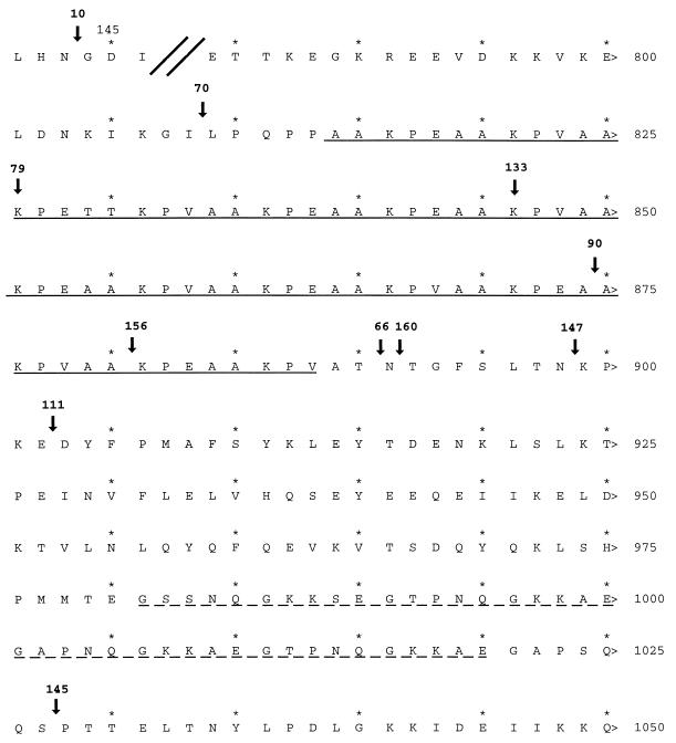
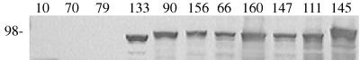
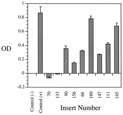
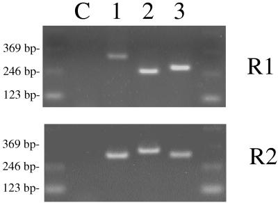
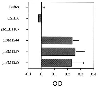
Mycoplasma hyopneumoniae colonizes the swine respiratory tract at the level of ciliated cells by attaching specifically to the cilium membrane. This interaction involves an adhesin called P97; the cilium binding activity of this protein was localized to the carboxy terminus, which included two repeat regions, R1 and R2 (T. Hsu, S. Artiushin, and F. C. Minion, J. Bacteriol. 179:1317-1323, 1997). To further delineate the molecular mechanisms of M. hyopneumoniae interactions with ciliated epithelium, we used a bank of transposon inserts in the cloned P97 gene to identify the site for cilium binding by testing the truncated gene products in an in vitro microtiter plate adherence assay. These studies showed that the cilium binding site was located in the AAKPV(E) repeat sequence of P97, referred to as the R1 repeat. For functional binding, at least seven AAKPV(E) repeats were required. The adherence-blocking monoclonal antibody F1B6 also recognized this region but required fewer AAKPV(E) repeats for recognition. We then constructed R1 region-lacZ gene fusions and used the resulting R1 repeat-beta-galactosidase fusion proteins in an in vitro assay to confirm the role of R1 in cilium binding. A comparison of the R1 regions of M. hyopneumoniae strains displaying variation in cilium adherence failed to identify changes that could account for the differences in adherence shown by the strains. Thus, we concluded that other proteins, in addition to P97, must be involved in cilium adherence, possibly in combination with P97.


Figures

FIG. 1
FIG. 1
Locations of Tn1000 insertions used to identify the MAb F1B6 binding epitope and the cilium binding site of P97. Shown is the translated amino acid sequence (accession no. U50901); amino acid positions are indicated by numbers at the right. Arrows indicate transposon insertion locations. The transposon insert number is given above each arrow. Insertion 10 is located upstream between amino acid positions 143 and 144. The solid underline indicates R1; the broken underline indicates R2.
FIG. 2
FIG. 2
Mapping the P97 F1B6 MAb binding epitope by immunoblotting using Tn1000 insertions in pISM2159. Whole-cell antigens were prepared from recombinant E. coli as described in Materials and Methods. The proteins were resolved on an SDS–10% polyacrylamide gel (3 μg per lane), and the resulting blot was developed with MAb F1B6. Each lane number indicates the number of the Tn1000 insertion (Fig. 1). The molecular mass given on the left in kilodaltons was determined from molecular weight markers (Bio-Rad). P97 does not undergo posttranslational cleavage in E. coli and would normally migrate as a 125-kDa protein, but the Tn1000 insertion results in truncation of the protein at the point of insertion.
FIG. 3
FIG. 3
Microtiter plate adherence assay analysis of Tn1000 insertions in P97. Lysates were prepared from ISM612 (pISM2159::Tn1000) strains and tested in the microtiter plate adherence assay as described in Materials and Methods. The data are presented as mean optical density (OD) ± standard error of triplicate wells. Numbers refer to positions of Tn1000 insertions in the P97 DNA sequence and correspond to lane numbers in Fig. 2. The positive control contains nonmutated pISM2159, and the negative control is lysate from ISM612 containing vector only.
FIG. 4
FIG. 4
Analysis of size variation in the P97 repeat regions by PCR analysis. The bands represent PCR products produced from template DNA from different M. hyopneumoniae strains. Lane C, no template DNA control; lane 1, strain 232A (adherent); lane 2, strain J (nonadherent); lane 3, strain 144L (adherent). R1 and R2 were amplified with primer sets TH120-TH121 and TH122-TH123, respectively.
FIG. 5
FIG. 5
Cilium binding activity of β-galactosidase fusion proteins containing R1 regions from different M. hyopneumoniae strains. The assay was performed as described previously (6) except that antibodies were omitted and o-nitrophenyl galactopyranoside was substituted as the substrate (9). Twenty micrograms of protein containing 28 U (pISM1244), 23 U (pISM1257), or 26 U (pISM1258) of β-galactosidase activity from E. coli lysates was added to each well, and the plates were incubated at 37°C for 90 min to allow binding of the fusion proteins to cilia. The control with pMLB1107 contained 44 U of activity. The data represent the mean optical density (OD) and standard deviation of six independent assays.

References

    1. Berman, M. Unpublished data.
    1. Dallo S F, Su C J, Horton J R, Baseman J B. Identification of P1 gene domain containing epitope(s) mediating Mycoplasma pneumoniae cytadherence. J Exp Med. 1988;167:718–723. - PMC - PubMed
    1. Gerstenecker B, Jacobs E. Topological mapping of the P1-adhesin of Mycoplasma pneumoniae with adherence-inhibiting monoclonal antibodies. J Gen Microbiol. 1990;136:471–476. - PubMed
    1. Guyer M S. Uses of transposon γδ in the analysis of cloned genes. Methods Enzymol. 1983;101:362–369. - PubMed
    1. Hanahan D. Studies on transformation of Escherichia coli with plasmids. J Mol Biol. 1983;166:557–580. - PubMed

Publication types

LinkOut - more resources