

Characterization of Clostridium botulinum type B neurotoxin associated with infant botulism in japan
- PMID: 9746583
- PMCID: PMC108594
- DOI: 10.1128/IAI.66.10.4811-4816.1998
Characterization of Clostridium botulinum type B neurotoxin associated with infant botulism in japan
Abstract
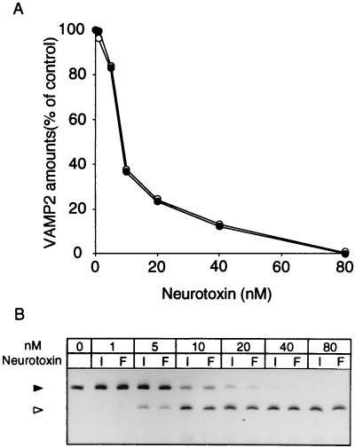
The neurotoxin of strain 111 (111/NT) associated with type B infant botulism showed antigenic and biological properties different from that (Okra/NT) produced by a food-borne botulism-related strain, Okra. The specific toxicity of 111/NT was found to be about 10 times lower than that of Okra/NT. The monoclonal antibodies recognizing the light chain cross-reacted with both neurotoxins, whereas most of the antibodies recognizing the carboxyl-terminal half of the heavy chain of Okra/NT did not react to 111/NT. Binding experiments with rat brain synaptosomes revealed that 125I-labeled 111/NT bound to a single binding site with a dissociation constant (Kd) of 2.5 nM; the value was rather lower than that (0.42 nM) of 125I-Okra/NT for the high-affinity binding site. In the lipid vesicles reconstituted with ganglioside GT1b, 125I-Okra/NT interacted with the amino-terminal domain of synaptotagmin 1 (Stg1N) or synaptotagmin 2 (Stg2N), fused with the maltose-binding protein, in the same manner as the respective full-length synaptotagmins, and the Kd values accorded with those of the low- and high-affinity binding sites in synaptosomes. However, 125I-111/NT only exhibited a low capacity for binding to the lipid vesicles containing Stg2N, but not Stg1N, in the presence of ganglioside GT1b. Moreover, synaptobrevin-2, an intracellular target protein, was digested to the same extent by the light chains of both neurotoxins in a concentration-dependent manner. These findings indicate that the 111/NT molecule possesses the receptor-recognition site structurally different from Okra/NT, probably causing a decreased specific toxicity.
Figures
References
-
- Arnon S S. Infant botulism. Annu Rev Med. 1981;31:541–560. - PubMed
-
- Arnon S S. Infant botulism: anticipating the second decade. J Infect Dis. 1986;154:201–205. - PubMed
-
- Bradford M M. A rapid and sensitive method for the quantitation of microgram quantities of protein utilizing the principle of protein-dye binding. Anal Biochem. 1976;72:248–254. - PubMed
-
- Brose N, Petrenko A G, Südhof T C, Jahn R. Synaptotagmin: a calcium sensor on the synaptic vesicle surface. Science. 1992;256:1021–1025. - PubMed
-
- Dodds K L. Worldwide incidence and ecology of infant botulism. In: Hauschild A H W, Dodds K L, editors. Clostridium botulinum: ecology and control in foods. New York, N.Y: Marcel Dekker, Inc.; 1992. pp. 105–117.
Publication types
MeSH terms
Substances
LinkOut - more resources
Full Text Sources
Other Literature Sources
Medical
Research Materials
