. 1998 Oct;66(10):4856-66.
doi: 10.1128/IAI.66.10.4856-4866.1998.

Experimental infection of Mongolian gerbils with wild-type and mutant Helicobacter pylori strains

Affiliations

Experimental infection of Mongolian gerbils with wild-type and mutant Helicobacter pylori strains

H P Wirth et al. Infect Immun. 1998 Oct.

Abstract

Experimental Helicobacter pylori infection was studied in Mongolian gerbils with fresh human isolates that carry or do not carry cagA (cagA-positive or cagA-negative, respectively), multiply passaged laboratory strains, wild-type strain G1.1, or isogenic ureA, cagA, or vacA mutants of G1.1. Animals were sacrificed 1 to 32 weeks after challenge, the stomach was removed from each animal for quantitative culture, urease test, and histologic testing, and blood was collected for antibody determinations. No colonization occurred after >/=20 in vitro passages of wild-type strain G1.1 or with the ureA mutant of G1.1. In contrast, infection occurred in animals challenged with wild-type G1.1 (99 of 101 animals) or the cagA (25 of 25) or vacA (25 of 29) mutant of G1.1. Infection with G1.1 persisted for at least 8 months. All 15 animals challenged with any of three fresh human cagA-positive isolates became infected, in contrast to only 6 (23%) of 26 animals challenged with one of four fresh human cagA-negative isolates (P < 0.001). Similar to infection in humans, H. pylori colonization of gerbils induced gastric inflammation and a systemic antibody response to H. pylori antigens. These data confirm the utility of gerbils as an animal model of H. pylori infection and indicate the importance of bacterial strain characteristics for successful infection.


Figures

FIG. 1
FIG. 1
Gastric colonization of Mongolian gerbils infected with H. pylori wild-type or mutant strains. Solid boxes, G1.1WT; open circles, cagA mutant; open triangles, vacA mutant. Animals were sacrificed 1 to 32 weeks after bacterial inoculation, and the number of CFU per stomach was determined by quantitative culture, as described in the text. The numbers of animals included are shown in Table 1. Results are expressed as mean and SEM for the infected animals.
FIG. 2
FIG. 2
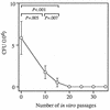
Effect of in vitro passage of H. pylori G1.1 on gastric colonization of Mongolian gerbils. A total of 5 or 10 animals in each group were challenged with strain G1.1 after 0 to 35 in vitro passages, and the number of CFU per stomach was determined by quantitative culture 1 and 2 weeks after challenge, as described in the text. Results are expressed as mean CFU per stomach of the originally challenged animals of the 1- and 2-week determinations and SEM. Student’s t test was used to calculate significance.
FIG. 3
FIG. 3
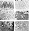
Histologic findings in the gastric antrum mucosa in uninfected (A and B) or H. pylori G1-1-infected (C to I) Mongolian gerbils. (A and B) Neither inflammatory infiltration (A) nor silver-stainable spiral microorganisms (B) are visible in a 10-week-old uninfected Mongolian gerbil challenged three times with brucella broth alone 2 weeks before sacrifice. The arrows in panel B indicate a narrow band of amorphous material at the mucosal surface, which was present equally in H. pylori-infected and uninfected animals. (C) The mucosa of an animal infected for 4 weeks shows a mild polymorphonuclear infiltrate with minimal cryptitis (neutrophils in glandular lumina) and mild chronic inflammation. (D) Mucosal erosion (arrow) where the superficial mucosa is focally infiltrated by polymorphonuclear inflammatory cells in an animal infected for 6 weeks. (E) Ulcer in a gerbil infected for 8 weeks. The surface epithelium is interrupted (arrows) by the ulceration, and the wall is heavily infiltrated by inflammatory cells. The muscularis mucosae is indicated by a star. (F) Same gerbil as in panel E. Silver-stainable dark brown curved or spiral-shaped microorganisms are visible in the ulcer floor and in glandular lumina at the ulcer edges. (G) Transmural inflammation in an animal infected for 8 weeks, involving the gastric wall from the mucosa (right) to the serosa (left). The muscularis mucosae is indicated by a star. (H) Mucosal infiltration of an animal infected for 32 weeks with focal replacement of gastric glands. (I) Antral lymphoid aggregates, some with germinal centers, were found after 4 weeks of infection in animals with dense mucosal (top) inflammation, which could extend beyond the muscularis mucosae (bottom; arrows), as in this gerbil after 8 weeks of infection. Magnifications: ×150 (A), ×195 (B), ×194 (C), ×195 (D), ×59 (E), ×208 (F), ×59 (G), ×91 (H), ×65 (I). Hematoxylin and eosin stain (A, C to E, and G to I); silver stain (B and F).
FIG. 3
FIG. 3
Histologic findings in the gastric antrum mucosa in uninfected (A and B) or H. pylori G1-1-infected (C to I) Mongolian gerbils. (A and B) Neither inflammatory infiltration (A) nor silver-stainable spiral microorganisms (B) are visible in a 10-week-old uninfected Mongolian gerbil challenged three times with brucella broth alone 2 weeks before sacrifice. The arrows in panel B indicate a narrow band of amorphous material at the mucosal surface, which was present equally in H. pylori-infected and uninfected animals. (C) The mucosa of an animal infected for 4 weeks shows a mild polymorphonuclear infiltrate with minimal cryptitis (neutrophils in glandular lumina) and mild chronic inflammation. (D) Mucosal erosion (arrow) where the superficial mucosa is focally infiltrated by polymorphonuclear inflammatory cells in an animal infected for 6 weeks. (E) Ulcer in a gerbil infected for 8 weeks. The surface epithelium is interrupted (arrows) by the ulceration, and the wall is heavily infiltrated by inflammatory cells. The muscularis mucosae is indicated by a star. (F) Same gerbil as in panel E. Silver-stainable dark brown curved or spiral-shaped microorganisms are visible in the ulcer floor and in glandular lumina at the ulcer edges. (G) Transmural inflammation in an animal infected for 8 weeks, involving the gastric wall from the mucosa (right) to the serosa (left). The muscularis mucosae is indicated by a star. (H) Mucosal infiltration of an animal infected for 32 weeks with focal replacement of gastric glands. (I) Antral lymphoid aggregates, some with germinal centers, were found after 4 weeks of infection in animals with dense mucosal (top) inflammation, which could extend beyond the muscularis mucosae (bottom; arrows), as in this gerbil after 8 weeks of infection. Magnifications: ×150 (A), ×195 (B), ×194 (C), ×195 (D), ×59 (E), ×208 (F), ×59 (G), ×91 (H), ×65 (I). Hematoxylin and eosin stain (A, C to E, and G to I); silver stain (B and F).
FIG. 4
FIG. 4
Evolution of gastric inflammation and colonization over time in Mongolian gerbils infected with H. pylori G1.1. Parameters were determined by histologic testing as described in the text, and scored from 0 to 3. Solid triangles, chronic inflammation; open triangles, acute inflammation. Upright and downward triangles (solid or open) represent superficial and deep (beyond the lamina muscularis mucosae) inflammation, respectively. Curved organisms detectable by silver staining in the glandular lumina are represented as open boxes. The numbers of animals included are shown in Table 1, except for weeks 2 and 3, when only three animals for each time were evaluated histologically. The results shown are mean values of the histologic scores and SEM.
FIG. 5
FIG. 5
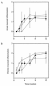
Comparison of gastric antrum inflammation in gerbils infected with G1.1WT and mutant strains. Acute (A) and chronic (B) superficial mucosal inflammation of animals after 1 to 12 weeks of infection with G1.1WT (solid squares), G1.1C (circles), or G1.1V (triangles) were determined by histologic testing, as described in the text. The results are expressed as mean histologic scores and SEM. The numbers of animals included are shown in Table 1. None of the differences was statistically significant.
FIG. 6
FIG. 6
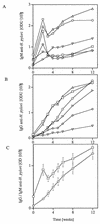
Variability of the serum antibody response to H. pylori in six representative infected gerbils during the first 12 weeks following challenge with strain G1.1. (A) IgM. (B) IgG. Blood specimens were obtained prechallenge, weekly from weeks 2 to 6, and at 8 and 12 weeks. Antibody levels in serum were determined by ELISA with a sonicate of G1.1 cells as the antigen. The results shown for each gerbil represent the mean of two duplicate determinations. The symbols for each gerbil are the same in both panels. (C) Mean values for IgG (circles) or IgM (squares) and SEM in 21 animals.

Similar articles

Cited by

References

    1. Abraham D, Weiner D J, Farrell J P. Cellular and humoral immune responses of jirds resistant to Dipetalonema viteae infection. Infect Immun. 1986;52:742–747. - PMC - PubMed
    1. Akopyanz N, Bukanov N O, Westblom T U, Kresovich S, Berg D E. DNA diversity among clinical isolates of Helicobacter pylori detected by PCR-based RAPD fingerprinting. Nucleic Acids Res. 1992;20:5137–5142. - PMC - PubMed
    1. Andrutis K A, Fox J G, Schauer D B, Marini R P, Murphy J C, Yan L, Solnick J V. Inability of an isogenic urease-negative mutant strain of Helicobacter mustelae to colonize the ferret stomach. Infect Immun. 1995;63:3722–3725. - PMC - PubMed
    1. Andrutis K A, Fox J G, Schauer D B, Marini R P, Li X, Yan L, Josenhans C, Suerbaum S. Infection of the ferret stomach by isogenic flagellar mutant strains of Helicobacter mustelae. Infect Immun. 1997;65:1962–1966. - PMC - PubMed
    1. Bartizal K F, Wostmann B S, Wagner M. Distribution and effects of a defined six-member murine-derived microflora in gnotobiotic gerbils. Appl Environ Microbiol. 1984;47:746–751. - PMC - PubMed

Publication types

MeSH terms