Comparative Study
. 1998 Oct;66(10):4875-83.
doi: 10.1128/IAI.66.10.4875-4883.1998.

Borrelia burgdorferi and interleukin-1 promote the transendothelial migration of monocytes in vitro by different mechanisms

Affiliations
Comparative Study

Borrelia burgdorferi and interleukin-1 promote the transendothelial migration of monocytes in vitro by different mechanisms

M J Burns et al. Infect Immun. 1998 Oct.

Abstract

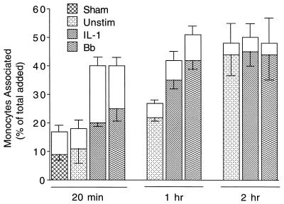
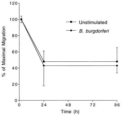
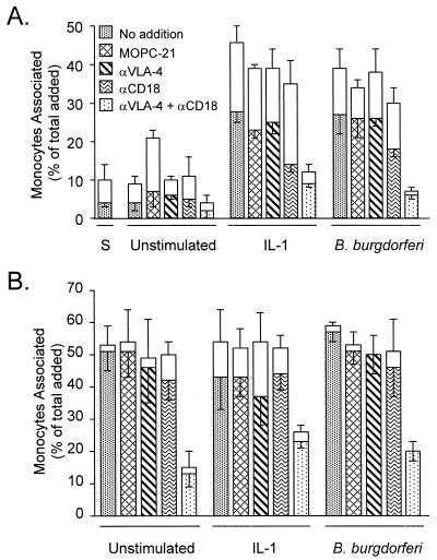
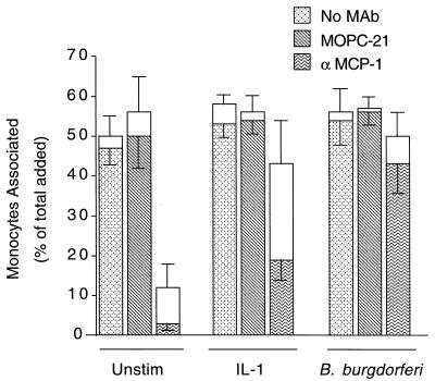
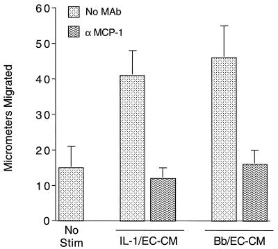
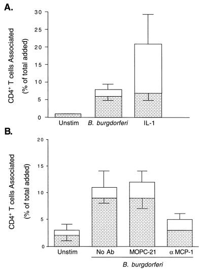
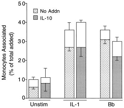
A prominent feature of Lyme disease is the perivascular accumulation of mononuclear leukocytes. Incubation of human umbilical vein endothelial cells (HUVEC) cultured on amniotic tissue with either interleukin-1 (IL-1) or Borrelia burgdorferi, the spirochetal agent of Lyme disease, increased the rate at which human monocytes migrated across the endothelial monolayers. Very late antigen 4 (VLA-4) and CD11/CD18 integrins mediated migration of monocytes across HUVEC exposed to either B. burgdorferi or IL-1 in similar manners. Neutralizing antibodies to the chemokine monocyte chemoattractant protein 1 (MCP-1) inhibited the migration of monocytes across unstimulated, IL-1-treated, or B. burgdorferi-stimulated HUVEC by 91% +/- 3%, 65% +/- 2%, or 25% +/- 22%, respectively. Stimulation of HUVEC with B. burgdorferi also promoted a 6-fold +/- 2-fold increase in the migration of human CD4(+) T lymphocytes. Although MCP-1 played only a limited role in the migration of monocytes across B. burgdorferi-treated HUVEC, migration of CD4(+) T lymphocytes across HUVEC exposed to spirochetes was highly dependent on this chemokine. The anti-inflammatory cytokine IL-10 reduced both migration of monocytes and endothelial production of MCP-1 in response to B. burgdorferi by approximately 50%, yet IL-10 inhibited neither migration nor secretion of MCP-1 when HUVEC were stimulated with IL-1. Our results suggest that activation of endothelium by B. burgdorferi may contribute to formation of the chronic inflammatory infiltrates associated with Lyme disease. The transendothelial migration of monocytes that is induced by B. burgdorferi is significantly less dependent on MCP-1 than is migration induced by IL-1. Selective inhibition by IL-10 further indicates that B. burgdorferi and IL-1 employ distinct mechanisms to activate endothelial cells.


Figures

FIG. 1
FIG. 1
Time course of the migration of monocytes across HUVEC stimulated with B. burgdorferi or IL-1. Monocytes were incubated for indicated times with HUVEC-amnion cultures that had been pretreated for 8 h with either control medium (Unstim), a sham preparation, 5 U of IL-1 per ml, or B. burgdorferi (Bb) at a ratio of 10 Bb/EC. Transendothelial migration was assessed as described in Materials and Methods. The total height of each bar represents the number of monocytes associated with each culture as a percentage of the total number added. The lower (patterned) portion of each bar represents the percentage that migrated beneath the endothelium; the upper (unfilled) portion represents the percentage adherent to the apical surface. Bars represent the means ± SD of three to four replicate samples. This experiment was repeated twice with similar results.
FIG. 2
FIG. 2
B. burgdorferi does not affect the rate at which monocytes exit from HUVEC-amnion cultures. HUVEC-amnion cultures were either left unstimulated or pretreated for 8 h with B. burgdorferi N40 at a ratio of 100 Bb/EC. Monocytes were then added to the cultures for 2 h. Cultures were washed and either fixed immediately or incubated for an additional 22 or 96 h. The number of monocytes remaining in the amniotic tissue was then determined as described in Materials and Methods. Data are presented as the percentage of monocytes that remained in the amniotic tissue relative to the number that had migrated after 2 h. Bars represent the means ± SD of four to five replicate samples. This experiment was repeated twice with similar results.
FIG. 3
FIG. 3
VLA-4 and CD11/CD18 integrins mediate the migration of monocytes across endothelium stimulated with B. burgdorferi or IL-1. Monocytes were suspended in medium containing either no addition, 50 μg of isotype-matched control MAb (MOPC-21) per ml, 10 μg of HP1/2 (anti-VLA-4) per ml, 40 μg of TS1/18 (anti-CD18) per ml, or 10 μg of HP1/2 plus 40 μg of TS1/18 per ml. The monocytes were then incubated for 20 min (A) or 2 h (B) with HUVEC that were either unstimulated or pretreated with a sham preparation (S), 5 U of IL-1 per ml, or B. burgdorferi at a ratio of 10 Bb/EC. The legend to panel A also applies to panel B. Transendothelial migration was assessed as described in Materials and Methods. The total height of each bar represents the number of monocytes associated with each culture as a percentage of the total number added. The lower (patterned) portion of each bar represents the percentage that migrated beneath the endothelium; the upper (unfilled) portion represents the percentage adherent to the apical surface. Bars represent the means ± SD of three to four replicate samples.
FIG. 4
FIG. 4
Time- and dose-dependent production of MCP-1 by HUVEC in response to B. burgdorferi or IL-1. Conditioned media were collected from HUVEC incubated with medium alone (Unstim), sham preparations, 5 U of IL-1 per ml, or B. burgdorferi at a ratio of 1, 10 or 100 Bb/EC for the indicated times. Amounts of MCP-1 were measured by ELISA. Datum points represent the means ± SD of three replicate samples. This experiment was repeated once with similar results.
FIG. 5
FIG. 5
MCP-1 is not the major chemoattractant mediating the migration of monocytes across B. burgdorferi-stimulated HUVEC. HUVEC cultures, elevated on silicone supports, were incubated for 8 h with either control medium (Unstim), 5 U of IL-1 per ml, or B. burgdorferi at a ratio of 10 Bb/EC in the presence of no MAb, 20 μg of control MAb (MOPC-21) per ml, or 20 μg of neutralizing anti-MCP-1 MAb per ml. Subsequently added monocytes, resuspended in fresh media containing the same MAbs, were incubated with the cultures for 2 h, and transendothelial migration was assessed as described in Materials and Methods. The total height of each bar represents the number of monocytes associated with each culture as a percentage of the total number added. The lower portion of each bar represents the percentage that migrated beneath the endothelium; the upper portion represents the percentage adherent to the apical surface. Bars represent the means ± SD of three replicate samples.
FIG. 6
FIG. 6
MCP-1 is the major soluble chemoattractant for monocytes produced by HUVEC in response to both IL-1 and B. burgdorferi. Conditioned media were collected from HUVEC incubated with either 5 U of IL-1 per ml (IL-1/EC-CM) or B. burgdorferi at a ratio of 10 Bb/EC (Bb/EC-CM) for 24 h. IL-1/EC-CM and Bb/EC-CM were diluted 1:10 in M199 and were either left untreated (No MAb) or incubated with 10 μg of neutralizing anti-MCP-1 MAb per ml. Conditioned media or control medium (No Stim) were tested for chemotactic activity toward monocytes in Boyden chambers. Bars represent the means ± SD of three replicate samples.
FIG. 7
FIG. 7
CD4+ T cells migrate across B. burgdorferi-stimulated HUVEC in an MCP-1-dependent manner. CD4+ T cells were incubated for 2 h with HUVEC-amnion cultures that had been pretreated with either control medium (Unstim), 5 U of IL-1 per ml, or B. burgdorferi at a ratio of 10 Bb/EC (A). CD4+ T cells were incubated for 2 h with HUVEC-amnion cultures that had been pretreated with either control medium (Unstim) or B. burgdorferi at a ratio of 10 Bb/EC in the presence of no MAb, an isotype-matched control MAb (MOPC-21), or 20 μg of neutralizing anti-MCP-1 MAb per ml (B). Transendothelial migration was assessed as described in Materials and Methods. The total height of each bar represents the number of T cells associated with each culture as a percentage of the total number added. The lower portion of each bar represents the percentage that migrated beneath the endothelium; the upper portion represents the percentage adherent to the apical surface. Bars represent the means ± SD of four to five replicate samples.
FIG. 8
FIG. 8
IL-10 inhibits the migration of monocytes across HUVEC stimulated by B. burgdorferi but not IL-1. Monocytes were added for 20 min to HUVEC-amnion cultures that had been pretreated for 8 h with control medium (Unstim), 5 U of IL-1 per ml, or B. burgdorferi at a ratio of 10 Bb/EC in the absence (No Addn) or presence of 20 ng of IL-10 per ml. Transendothelial migration was assessed as described in Materials and Methods. The total height of each bar represents the number of monocytes associated with each culture as a percentage of the total added. The lower portion of each bar represents the percentage that migrated beneath the endothelium; the upper portion represents the percentage adherent to the apical surface. Bars represent the means ± SD of three to five replicate samples. This experiment yielded similar results when repeated once using both IL-1- and B. burgdorferi-stimulated samples and three additional times with B. burgdorferi-treated samples only.

Similar articles

Cited by

References

    1. Anonymous. Lyme disease—United States, 1996. Morbid Mortal Weekly Rep. 1997;46:531–535. - PubMed
    1. Baggiolini M, Dewald B, Moser B. Interleukin-8 and related chemotactic cytokines-CXC and CC chemokines. Adv Immunol. 1994;55:97–179. - PubMed
    1. Baggiolini M, Dewald B, Moser B. Human chemokines: an update. Annu Rev Immunol. 1997;15:675–705. - PubMed
    1. Baldwin A S., Jr The NF-κB and IκB proteins: new discoveries and insights. Annu Rev Immunol. 1996;14:649–683. - PubMed
    1. Barthold S W, Beck D S, Hansen G M, Terwilliger G A, Moody K D. Lyme borreliosis in selected strains and ages of laboratory mice. J Infect Dis. 1990;162:133–138. - PubMed

Publication types

MeSH terms