. 1998 Oct;66(10):4924-31.
doi: 10.1128/IAI.66.10.4924-4931.1998.

Cloning and molecular characterization of a cDNA clone coding for Trichomonas vaginalis alpha-actinin and intracellular localization of the protein

Affiliations

Cloning and molecular characterization of a cDNA clone coding for Trichomonas vaginalis alpha-actinin and intracellular localization of the protein

M F Addis et al. Infect Immun. 1998 Oct.

Abstract

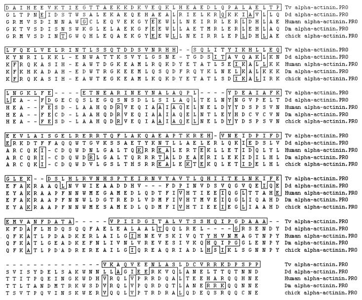
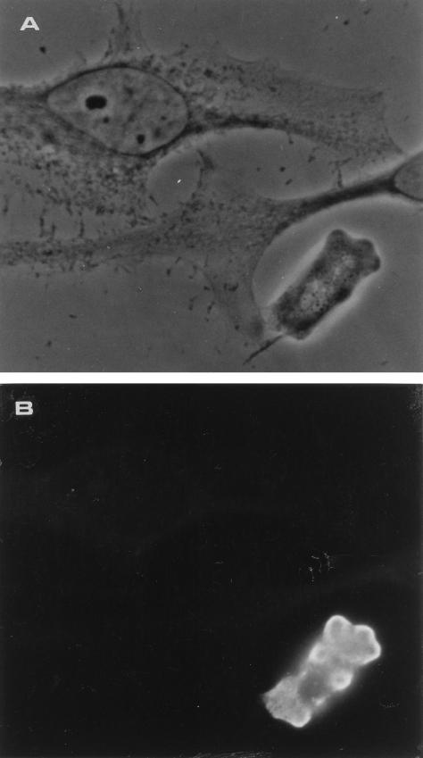
We have identified and sequenced a cDNA clone coding for Trichomonas vaginalis alpha-actinin. Analysis of the obtained sequence revealed that the 2,857-nucleotide-long cDNA contained an open reading frame encoding 849 amino acids which showed consistent homology with alpha-actinins of different species. Such homology was particularly significant in regions which have been reported to represent the actin-binding and Ca2+-binding domains in other alpha-actinins. The deduced protein was also characterized by the presence of a divergent central region thought to play a role in its high immunogenicity. A study of protein localization performed by immunofluorescence revealed that the protein is diffusely distributed throughout the T. vaginalis cytoplasm when the cell is pear shaped. When parasites adhere and transform into the amoeboid morphology, the protein is located only in areas close to the cytoplasmic membrane and colocalizes with actin. Concomitantly with transformation into the amoeboid morphology, alpha-actinin mRNA expression is upregulated.


Figures

FIG. 1
FIG. 1
(A) Deduced amino acid sequence of the T. vaginalis alpha-actinin cDNA. The actin-binding site and the EF-hand domain are shown in bold. The underlined amino acids represent the central, divergent region. (B) Homology between actin-binding site of T. vaginalis alpha-actinin and actin-binding sites of other alpha-actinins. Consensus indicates the most frequent amino acid at that position. Uppercase letters indicate that the amino acid is present in all peptides; lowercase letters indicate the most common amino acid. (C) Homology between EF-hand (Ca2+-chelating domain) of T. vaginalis alpha-actinin and EF-hands of other alpha-actinins. Consensus indicates the EF-hand consensus sequence (33). The calcium-chelating side chains are also shown (x, y, z-y-x, and -z). D, oxygen-containing side chain; L, hydrophobic side chain; Tv, T. vaginalis; Dd, D. discoideum; Drome, D. melanogaster.
FIG. 2
FIG. 2
Alignment of the alpha-actinin central region in amino acid sequences from T. vaginalis (Tv), D. discoideum (Dd), Homo sapiens (Human), D. melanogaster (Dm), and chick. Boxes represent the amino acids that match those in T. vaginalis alpha-actin exactly. The alignment was obtained by the Clustal method with the MegAlign program (Lasergene Suite; DnaStar Inc., Madison, Wis.).
FIG. 3
FIG. 3
(A) Phase-contrast micrograph of an amoeboid T. vaginalis organism coincubated with HeLa cells. (B) Immunofluorescence pattern of alpha-actinin in the corresponding sample. Cells were fixed and processed for immunofluorescence staining with anti–T. vaginalis alpha-actinin antibodies. These images show the lack of cross-reactivity between parasite and host alpha-actinins.
FIG. 4
FIG. 4
Distribution of alpha-actinin in different morphological forms of T. vaginalis. Living T. vaginalis organisms in suspension and adhering on coverslips were fixed and processed for immunofluorescence staining with anti–T. vaginalis alpha-actinin antibodies. (Right panels) Immunofluorescence patterns of alpha-actinin during different morphological stages of the parasites: A, pear-shaped form; D, fully amoeboid form; B and C, intermediate stages. (Left panel) Phase-contrast micrographs of the corresponding samples. This sequence of images shows the redistribution of alpha-actinin in the periphery of the microorganism following transformation into the amoeboid morphology.
FIG. 5
FIG. 5
Representative experiment showing a Northern blot and RT-PCR of total mRNA extracted from pear-shaped (lanes a) and amoeboid (lanes b) T. vaginalis organisms. (Northern blot) Panel 1, hybridization bands obtained with an actin probe; panel 2, hybridization bands obtained with an alpha-actinin probe. (RT-PCR) Panel 3, amplification bands obtained from RNA extracted from pear-shaped and amoeboid parasites with actin primers; panel 4, amplification bands obtained with alpha-actinin primers.

Similar articles

Cited by

References

    1. Addis M F, Rappelli P, Cappuccinelli P, Fiori P L. Extracellular release by Trichomonas vaginalis of a NADP+ dependent malic enzyme involved in pathogenicity. Microb Pathog. 1997;23:55–61. - PubMed
    1. Addis, M. F., et al. Unpublished data.
    1. Alderete J F, Pearlman E. Pathogenic Trichomonas vaginalis cytotoxicity to cell culture monolayers. Br J Vener Dis. 1984;60:99–105. - PMC - PubMed
    1. Altschul S F, Madden T L, Schaffer A A, Zhang J, Zhang Z, Miller W, Lipman D J. Gapped BLAST and PSI-BLAST: a new generation of protein database search programs. Nucleic Acids Res. 1997;25:3389–3402. - PMC - PubMed
    1. Arroyo R, Engbring J, Alderete J F. Molecular basis of host epithelial cell recognition by Trichomonas vaginalis. Mol Microbiol. 1992;6:853–862. - PubMed

Publication types

MeSH terms

Associated data