

The cell cycle-specific growth-inhibitory factor produced by Actinobacillus actinomycetemcomitans is a cytolethal distending toxin
- PMID: 9746611
- PMCID: PMC108622
- DOI: 10.1128/IAI.66.10.5008-5019.1998
The cell cycle-specific growth-inhibitory factor produced by Actinobacillus actinomycetemcomitans is a cytolethal distending toxin
Abstract
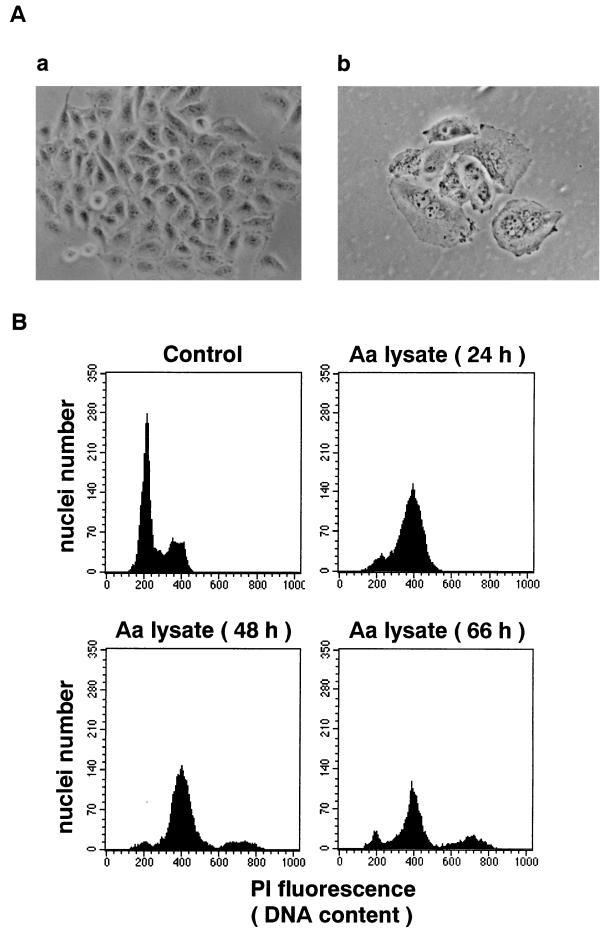
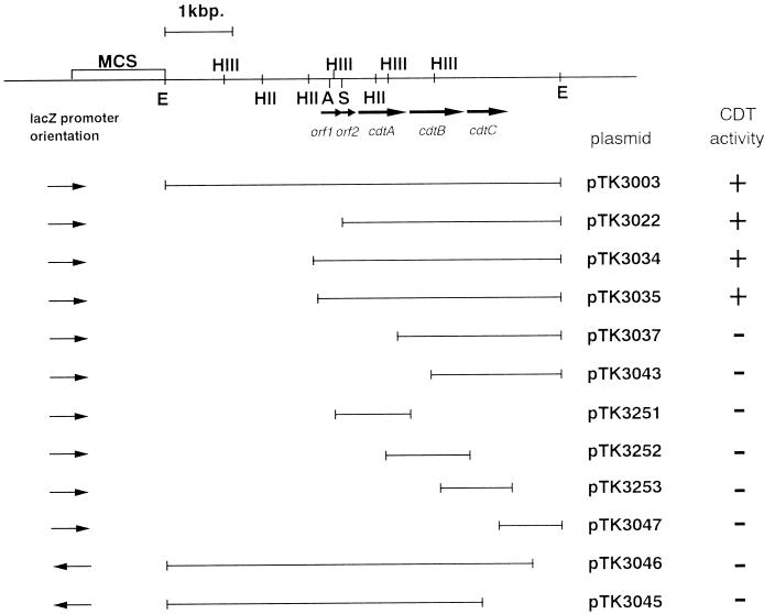
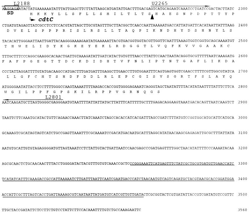
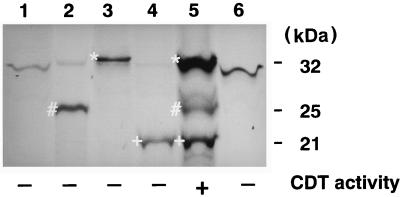
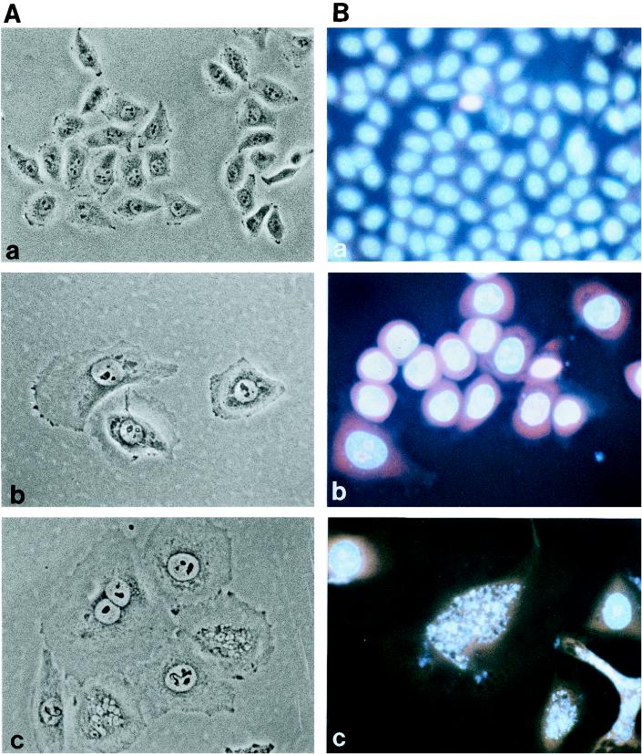
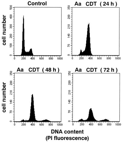
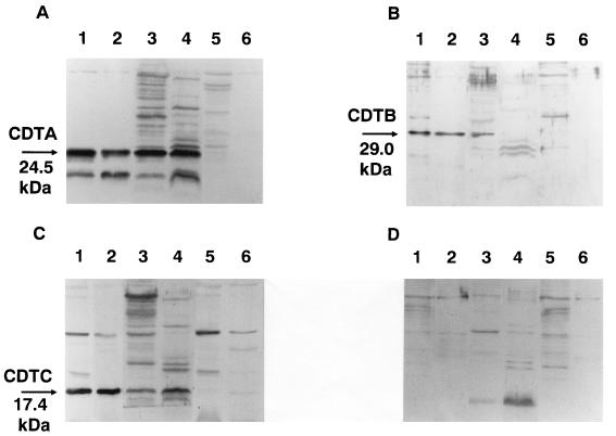
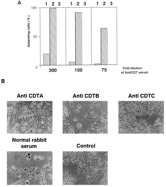
Actinobacillus actinomycetemcomitans has been shown to produce a soluble cytotoxic factor(s) distinct from leukotoxin. We have identified in A. actinomycetemcomitans Y4 a cluster of genes encoding a cytolethal distending toxin (CDT). This new member of the CDT family is similar to the CDT produced by Haemophilus ducreyi. The CDT from A. actinomycetemcomitans was produced in Escherichia coli and was able to induce cell distension, growth arrest in G2/M phase, nucleus swelling, and chromatin fragmentation in HeLa cells. The three proteins, CDTA, -B and -C, encoded by the cdt locus were all required for toxin activity. Antiserum raised against recombinant CDTC completely inhibited the cytotoxic activity of culture supernatant and cell homogenate fractions of A. actinomycetemcomitans Y4. These results strongly suggest that the CDT is responsible for the cytotoxic activity present in the culture supernatant and cell homogenate fractions of A. actinomycetemcomitans Y4. This CDT is a new putative virulence factor of A. actinomycetemcomitans and may play a role in the pathogenesis of periodontal diseases.
Figures

Similar articles
-
Expression of the cytolethal distending toxin (Cdt) operon in Actinobacillus actinomycetemcomitans: evidence that the CdtB protein is responsible for G2 arrest of the cell cycle in human T cells.J Immunol. 2000 Sep 1;165(5):2612-8. doi: 10.4049/jimmunol.165.5.2612. J Immunol. 2000. PMID: 10946289
-
Actinobacillus actinomycetemcomitans cytolethal distending toxin (Cdt): evidence that the holotoxin is composed of three subunits: CdtA, CdtB, and CdtC.J Immunol. 2004 Jan 1;172(1):410-7. doi: 10.4049/jimmunol.172.1.410. J Immunol. 2004. PMID: 14688349
-
Resistance of human periodontal ligament fibroblasts to the cytolethal distending toxin of Actinobacillus actinomycetemcomitans.J Periodontol. 2005 Jul;76(7):1189-201. doi: 10.1902/jop.2005.76.7.1189. J Periodontol. 2005. PMID: 16018764 Free PMC article.
-
The contribution of cytolethal distending toxin to bacterial pathogenesis.Crit Rev Microbiol. 2006 Oct-Dec;32(4):227-48. doi: 10.1080/10408410601023557. Crit Rev Microbiol. 2006. PMID: 17123907 Review.
-
The cytolethal distending toxin family.Trends Microbiol. 1999 Jul;7(7):292-7. doi: 10.1016/s0966-842x(99)01537-1. Trends Microbiol. 1999. PMID: 10390639 Review.
Cited by
-
A Mycobacterium ulcerans toxin, mycolactone, causes apoptosis in guinea pig ulcers and tissue culture cells.Infect Immun. 2000 Feb;68(2):877-83. doi: 10.1128/IAI.68.2.877-883.2000. Infect Immun. 2000. PMID: 10639458 Free PMC article.
-
CdtA, CdtB, and CdtC form a tripartite complex that is required for cytolethal distending toxin activity.Infect Immun. 2001 Jul;69(7):4358-65. doi: 10.1128/IAI.69.7.4358-4365.2001. Infect Immun. 2001. PMID: 11401974 Free PMC article.
-
Cytolethal Distending Toxin From Campylobacter jejuni Requires the Cytoskeleton for Toxic Activity.Jundishapur J Microbiol. 2016 Sep 18;9(10):e35591. doi: 10.5812/jjm.35591. eCollection 2016 Oct. Jundishapur J Microbiol. 2016. PMID: 27942359 Free PMC article.
-
The Enterobacterial Genotoxins: Cytolethal Distending Toxin and Colibactin.EcoSal Plus. 2016 Jul;7(1):10.1128/ecosalplus.ESP-0008-2016. doi: 10.1128/ecosalplus.ESP-0008-2016. EcoSal Plus. 2016. PMID: 27419387 Free PMC article. Review.
-
Variation of loop sequence alters stability of cytolethal distending toxin (CDT): crystal structure of CDT from Actinobacillus actinomycetemcomitans.Protein Sci. 2006 Feb;15(2):362-72. doi: 10.1110/ps.051790506. Protein Sci. 2006. PMID: 16434747 Free PMC article.
References
-
- Billington S J, Sinistraj M, Cheetham M, Ayres B F, Moses E K, Katz M E, Rood J I. Identification of a native Dichelobacter nodosus plasmid and implications for the evolution of the vap regions. Gene. 1996;172:111–116. - PubMed
-
- Bouzari S, Vatsala B R, Varghese A. In vitro adherence property of cytolethal distending toxin (CLDT) producing EPEC strains and effect of the toxin on rabbit intestine. Microb Pathog. 1992;12:153–157. - PubMed
-
- Braun V, Wu H C. Lipoproteins, structure, function, biosynthesis and model for protein export. New Compr Biochem. 1994;27:319–341.
-
- Bullock W O, Fernandez J M, Short J M. XL1-Blue: a high efficiency plasmid transforming recA Escherichia coli strain with beta-galactosidase selection. BioTechniques. 1987;5:376–379.
Publication types
MeSH terms
Substances
Associated data
- Actions
LinkOut - more resources
Full Text Sources
Molecular Biology Databases
