

Acquisition of iron by Gardnerella vaginalis
- PMID: 9746616
- PMCID: PMC108627
- DOI: 10.1128/IAI.66.10.5041-5047.1998
Acquisition of iron by Gardnerella vaginalis
Abstract
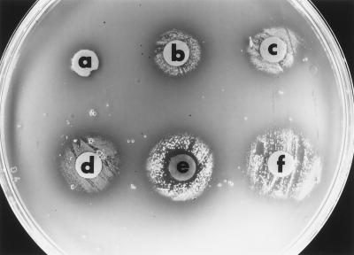
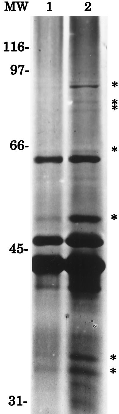
Six Gardnerella vaginalis strains were examined for the ability to utilize various iron-containing compounds as iron sources. In a plate bioassay, all six strains acquired iron from ferrous chloride, ferric chloride, ferrous sulfate, ferric ammonium citrate, ferrous ammonium sulfate, bovine and equine hemin, bovine catalase, and equine, bovine, rabbit, and human hemoglobin. All six strains also acquired iron from human lactoferrin, but not from human transferrin, as determined by a liquid broth growth assay. Siderophore production was detected in eight G. vaginalis strains by the chrome azurol S universal chemical assay. Sodium dodecyl sulfate-polyacrylamide gel electrophoresis of the cytoplasmic membrane proteins isolated from G. vaginalis 594 grown under iron-replete and iron-restricted conditions revealed several iron-regulated proteins ranging in molecular mass from 33 to 94 kDa. These results indicate that G. vaginalis may acquire iron from iron salts and host iron compounds.
Figures
Similar articles
-
Binding of heme by Gardnerella vaginalis.J Basic Microbiol. 2001;41(1):37-43. doi: 10.1002/1521-4028(200103)41:1<37::AID-JOBM37>3.0.CO;2-W. J Basic Microbiol. 2001. PMID: 11314245
-
Identification of a human lactoferrin-binding protein in Gardnerella vaginalis.Infect Immun. 2000 Jun;68(6):3443-7. doi: 10.1128/IAI.68.6.3443-3447.2000. Infect Immun. 2000. PMID: 10816496 Free PMC article.
-
Binding of catalase by Gardnerella vaginalis.FEMS Microbiol Lett. 2000 Sep 15;190(2):191-4. doi: 10.1111/j.1574-6968.2000.tb09285.x. FEMS Microbiol Lett. 2000. PMID: 11034278
-
Identification of a Gardnerella vaginalis hemoglobin-binding protein.Curr Microbiol. 2001 Jan;42(1):49-52. doi: 10.1007/s002840010177. Curr Microbiol. 2001. PMID: 11116397
-
Iron metabolism in pathogenic bacteria.Annu Rev Microbiol. 2000;54:881-941. doi: 10.1146/annurev.micro.54.1.881. Annu Rev Microbiol. 2000. PMID: 11018148 Review.
Cited by
-
Antimicrobial activity of bovine lactoferrin against Gardnerella species clinical isolates.Front Microbiol. 2022 Sep 8;13:1000822. doi: 10.3389/fmicb.2022.1000822. eCollection 2022. Front Microbiol. 2022. PMID: 36419418 Free PMC article.
-
Incubation period and risk factors support sexual transmission of bacterial vaginosis in women who have sex with women.Sex Transm Infect. 2019 Nov;95(7):511-515. doi: 10.1136/sextrans-2018-053824. Epub 2019 Mar 14. Sex Transm Infect. 2019. PMID: 30872415 Free PMC article.
-
Prevalence and clinical correlates of Gardnerella spp., Fannyhessea vaginae, Lactobacillus crispatus and L. iners in pregnant women in Bukavu, Democratic Republic of the Congo.Front Cell Infect Microbiol. 2025 Jan 17;14:1514884. doi: 10.3389/fcimb.2024.1514884. eCollection 2024. Front Cell Infect Microbiol. 2025. PMID: 39897482 Free PMC article.
-
Association of Female Genital Schistosomiasis With the Cervicovaginal Microbiota and Sexually Transmitted Infections in Zambian Women.Open Forum Infect Dis. 2021 Aug 22;8(9):ofab438. doi: 10.1093/ofid/ofab438. eCollection 2021 Sep. Open Forum Infect Dis. 2021. PMID: 34557562 Free PMC article.
-
Healthy Vaginal Microbiota and Influence of Probiotics Across the Female Life Span.Front Microbiol. 2022 Apr 8;13:819958. doi: 10.3389/fmicb.2022.819958. eCollection 2022. Front Microbiol. 2022. PMID: 35464937 Free PMC article. Review.
References
-
- Amsel R, Totten P A, Spiegel C A, Chen K C S, Eschenbach D, Holmes K K. Nonspecific vaginitis: diagnostic criteria and microbial and epidemiologic associations. Am J Med. 1983;74:14–22. - PubMed
-
- Boustouller Y L, Johnson A P, Taylor-Robinson D. Pili on Gardnerella vaginalis studied by electron microscopy. J Med Microbiol. 1987;23:327–329. - PubMed
Publication types
MeSH terms
Substances
LinkOut - more resources
Full Text Sources
Other Literature Sources
Medical
Molecular Biology Databases
