. 1998 Oct;180(19):5030-7.
doi: 10.1128/JB.180.19.5030-5037.1998.

New potential cell wall glucanases of Saccharomyces cerevisiae and their involvement in mating

Affiliations

New potential cell wall glucanases of Saccharomyces cerevisiae and their involvement in mating

C Cappellaro et al. J Bacteriol. 1998 Oct.

Abstract

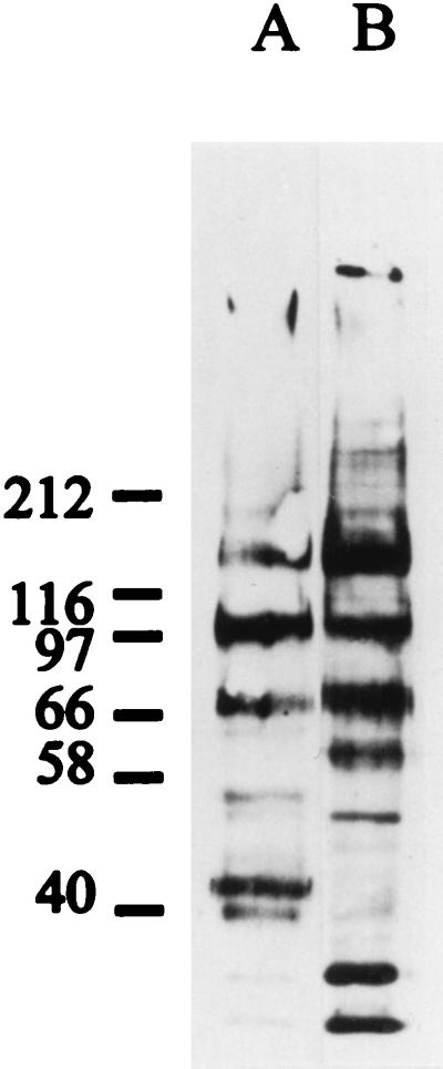
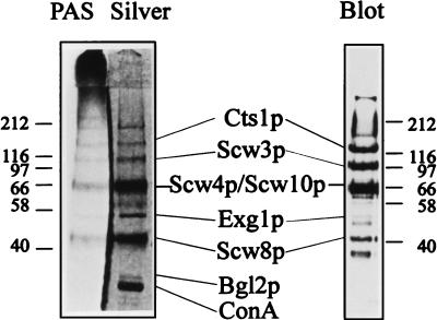
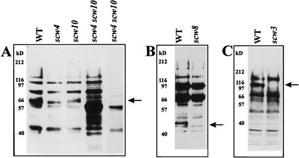
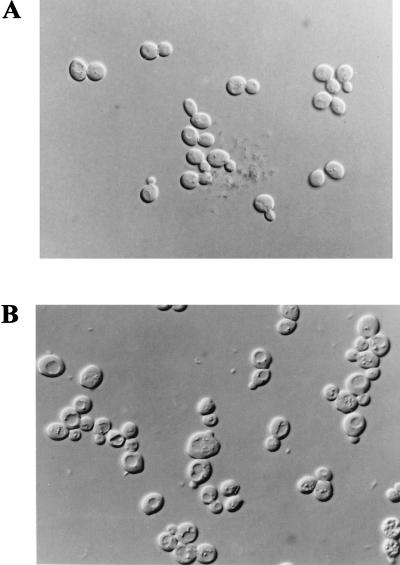
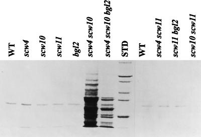
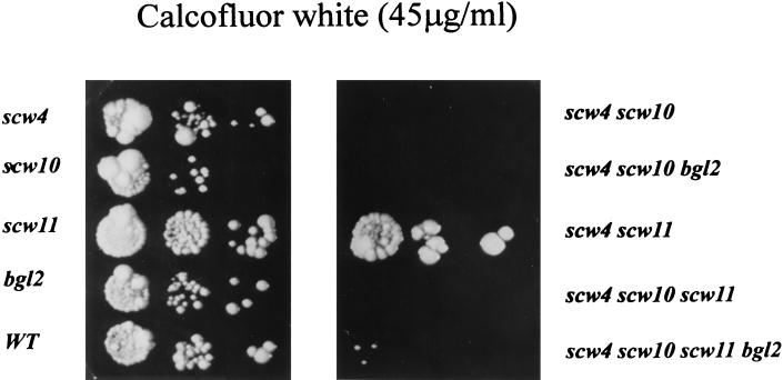
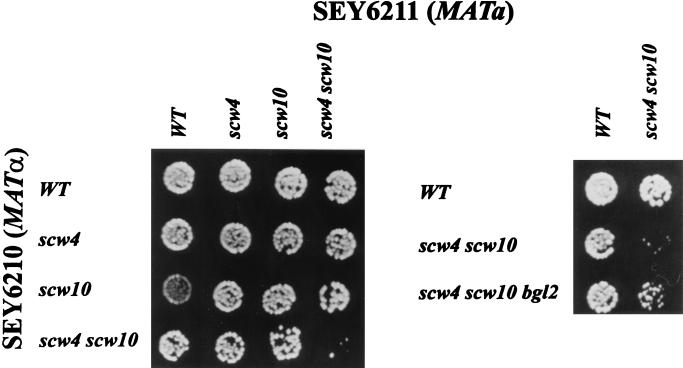
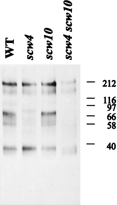
Biotinylation of intact Saccharomyces cerevisiae cells with a nonpermeant reagent (Sulfo-NHS-LC-Biotin) allowed the identification of seven cell wall proteins that were released from intact cells by dithiothreitol (DTT). By N-terminal sequencing, three of these proteins were identified as the known proteins beta-exoglucanase 1 (Exg1p), beta-endoglucanase (Bgl2p), and chitinase (Cts1p). One protein was related to the PIR protein family, whereas the remaining three (Scw3p, Scw4p, and Scw10p [for soluble cell wall proteins]) were found to be related to glucanases. Single knockouts of these three potential glucanases did not result in dramatic phenotypes. The double knockout of SCW4 and the homologous gene SCW10 resulted in slower growth, significantly increased release of proteins from intact cells by DTT, and highly decreased mating efficiency when these two genes were disrupted in both mating types. The synergistic behavior of the disruption of SCW4 and SCW10 was partly antagonized by the disruption of BGL2. The data are discussed in terms of a possible counterplay of transglucosidase and glucosidase activities.


Figures

FIG. 1
FIG. 1
Comparison of two extraction methods for Sulfo-NHS-LC-Biotin-labeled cell wall proteins. This blot was stained with a streptavidin-peroxidase conjugate. (A) Extraction of intact cells with DTT. An amount of extract equivalent to 4 ml of cells with an OD578 of 5 was subjected to SDS-PAGE. (B) Cell walls isolated from broken cells were extracted by heating in Laemmli sample buffer under reducing conditions (2% SDS, 5% β-mercaptoethanol). The equivalent of 200 μl of cells with an OD578 of 5 was applied. For the molecular weight standards used, see Materials and Methods. The values beside the gel are molecular sizes in kilodaltons.
FIG. 2
FIG. 2
Analysis of isolated proteins. (Left) Concentrated and ConA-purified DTT extract of intact cells stained for sugars (PAS) and protein (silver). (Right) Parallel extract isolated from biotinylated cells blotted and visualized with a streptavidin-peroxidase conjugate. The coincident patterns indicate that the purified materials are surface-exposed, glycosylated proteins. The names given resulted from N-terminal sequencing (Table 1). Cts1p, chitinase; Exg1p, exoglucanase 1; Bgl2p, β-endoglucanase 2; Scw8p, protein with an unknown function; Scw3p, Scw4p, and Scw10p, soluble cell wall proteins 3, 4, and 10 (see text). ConA is a contamination due to the purification procedure.
FIG. 3
FIG. 3
Scw4p and three related gene products are potential glucanases. Amino acid sequences of Scw4p (sequence 1), Scw10p (sequence 2), Scw11p (sequence 3), and Bgl2p (sequence 4) are compared. The Munich Information Center for Protein Sequences database was screened with the sequence of Scw4p (by using FASTA). The resulting ORFs were aligned by using the pileup function of The University of Wisconsin Genetics Computer Group package. ORFs are available from the Munich Information Center for Protein Sequences under the following accession numbers (PIR code): Scw4p, S64614; Scw10p, S53975; Scw11p, S64030, Bgl2p, A33499. Boldface letters are used when at least two amino acids are identical.
FIG. 4
FIG. 4
Grouping of known and potential glucanases. ORFs of gene products related to known glucanases were obtained from the Munich Information Center for Protein Sequences based on a FASTA search with the sequences of Exg1p, Scw4p, and Scw3p (S53916). Sequences were processed with the lineup function of the University of Wisconsin Genetics Computer Group package.
FIG. 5
FIG. 5
scw4, scw10, scw8 and scw3 knockout mutants. The corresponding mutant cells were labeled with Sulfo-NHS-LC-biotin and extracted with DTT (scw4, scw10, and scw8), or whole cells were heated in Laemmli sample buffer (scw3). For the molecular size standards, see Materials and Methods. The arrows mark gene products missing in the mutants. Lane 5 in panel A contains 1/10 of the material in lane 4. WT, wild type.
FIG. 6
FIG. 6
Morphology of wild-type (A) and scw4 scw10 double-mutant (B) cells.
FIG. 7
FIG. 7
Demonstration of cell wall lability. Log-phase cells were collected and extracted with DTT, and an aliquot was separated by SDS-PAGE and stained with Coomassie brilliant blue R-250. An amount corresponding to 6.6 ml of cells at an OD578 of 3.0 was applied to each lane of the gel. WT, wild type.
FIG. 8
FIG. 8
Synergistic effect of the scw4 and scw10 mutations on calcofluor white sensitivity. Five-microliter volumes of different dilutions (2 × 10⁵, 2 × 10⁴, and 2 × 10³ cells/ml) of stationary-phase cell cultures were dropped onto YPD plates containing 45 μg of calcofluor white and incubated for 5 days at 29°C. WT, wild type.
FIG. 9
FIG. 9
Requirement of Scw4p and Scw10p for mating. The mutants indicated in a Lys background (SEY 6210, MATα) were mated with mutants in an Ade background (SEY6211, MATa). Portions (104 cells) of stationary-phase cell cultures were mixed and dropped onto Lys Ade minimal-medium plates for detection of diploid (Lys+ Ade+) cells. WT, wild type.
FIG. 10
FIG. 10
Influence of Scw4p and Scw10p on the incorporation of covalently bound cell wall proteins. Cell wall proteins of the wild type (WT) and mutants were labeled by biotinylation (see Materials and Methods). Walls were purified, and the SDS-extractable proteins were extracted by Laemmli sample buffer. The remaining covalently attached proteins were then solubilized by laminarinase, subjected to electrophoresis, and blotted, and the blot was visualized by using a streptavidin-peroxidase conjugate. Each lane contains proteins obtained from cells corresponding to 25 U of OD600. The values on the right are molecular sizes in kilodaltons.

References

    1. Angiolella L, Facchin M, Stringaro A, Maras B, Simonetti N, Cassone A. Identification of a glucan-associated enolase as a main cell wall protein of Candida albicansand an indirect target of lipopeptide antimycotics. J Infect Dis. 1996;173:684–690. - PubMed
    1. Broach J R, Jones E W, Pringle J R, editors. The molecular and cellular biology of the yeast Saccharomyces. 1. Genome dynamics, protein synthesis and energetics. Plainview, N.Y: Cold Spring Harbor Laboratory Press; 1991.
    1. Cappellaro, C. Unpublished data.
    1. Casanova M, Lopez-Ribot J L, Martinez J P, Sentandreu R. Characterization of cell wall proteins from yeast and mycelial cells of Candida albicansby labeling with biotin: comparison with other techniques. Infect Immun. 1992;60:4898–4906. - PMC - PubMed
    1. Goffeau A, Barrell B G, Bussey H, Davis R W, Dujon B, et al. Life with 6000 genes. Science. 1996;274:546–567. - PubMed

Publication types