. 1998 Oct;180(19):5085-93.
doi: 10.1128/JB.180.19.5085-5093.1998.

Characterization of an A-factor-responsive repressor for amfR essential for onset of aerial mycelium formation in Streptomyces griseus

Affiliations

Characterization of an A-factor-responsive repressor for amfR essential for onset of aerial mycelium formation in Streptomyces griseus

K Ueda et al. J Bacteriol. 1998 Oct.

Abstract

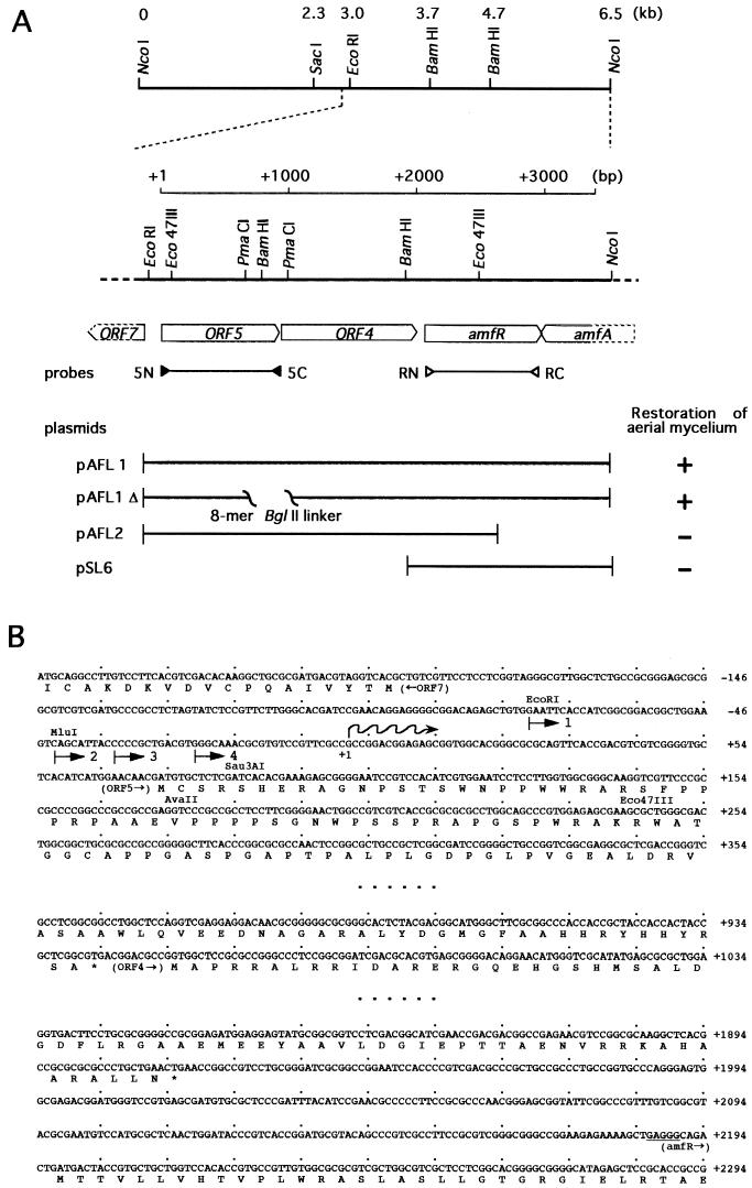
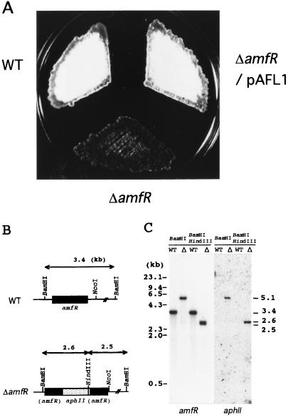
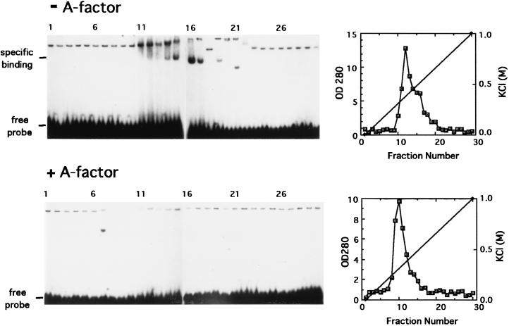
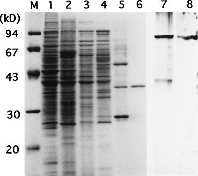
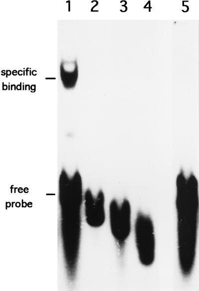
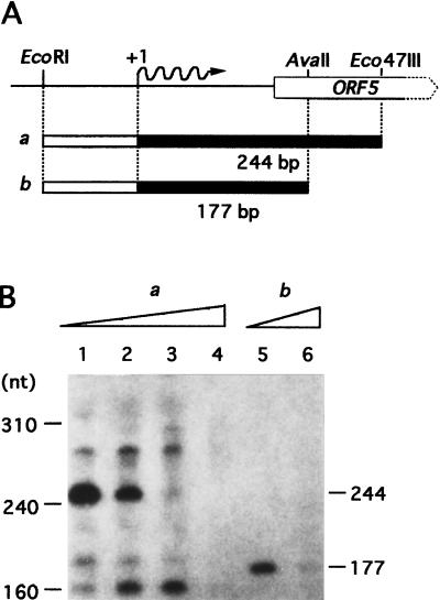
A-factor (2-isocapryloyl-3R-hydroxymethyl-gamma-butyrolactone) is essential for the initiation of aerial mycelium formation in Streptomyces griseus. amfR is one of the genes which, when cloned on a low-copy-number plasmid, suppresses the aerial mycelium-negative phenotype of an A-factor-deficient mutant of S. griseus. Disruption of the chromosomal amfR gene resulted in complete abolition of aerial mycelium formation, indicating that amfR is essential for the onset of morphogenesis. Cloning and nucleotide sequencing of the region upstream of amfR predicted an operon consisting of orf5, orf4, and amfR. Consistent with this idea, Northern blotting and S1 mapping analyses suggested that these three genes were cotranscribed mainly by a promoter (PORF5) in front of orf5. Furthermore, PORF5 was active only in the presence of A-factor, indicating that it is A-factor dependent. Gel mobility shift assays showed the presence of a protein (AdpB) able to bind PORF5 in the cell extract from an A-factor-deficient mutant but not from the wild-type strain. AdpB was purified to homogeneity and found to bind specifically to the region from -72 to -44 bp with respect to the transcriptional start point. Runoff transcriptional analysis of PORF5 with purified AdpB and an RNA polymerase complex isolated from vegetative mycelium showed that AdpB repressed the transcription in a concentration-dependent manner. It is thus apparent that AmfR as a switch for aerial mycelium formation and AdpB as a repressor for amfR are members in the A-factor regulatory cascade, leading to morphogenesis.


Figures

FIG. 1
FIG. 1
Restriction map of the 6.5-kb region containing orf5-orf4-amfR-amfA and nucleotide sequences of part of this region. (A) The directions and extents of the ORFs deduced from the nucleotide sequence are shown just below the restriction map. The positions of a pair of the primers used for probes for Northern hybridization (Fig. 3) are indicated. The fragments on a low-copy-number plasmid and their ability to restore aerial mycelium formation in S. griseus HH1 are shown. (B) The nucleotide sequences of the region including the promoter in front of orf5, the termination codon of orf5 and the initiation codon of orf4, and the termination codon of orf4 and the initiation codon of amfR. Probable ribosome-binding sites for orf5 and amfR are underlined. The transcriptional start point is indicated as +1. Numbers with an arrow indicate the probes used for the gel mobility shift assay (Fig. 4).
FIG. 2
FIG. 2
Phenotypes of the amfR disruptant (A), schematic representation of the disrupted amfR (B), and Southern hybridization analysis of the chromosome of the amfR disruptant (C). (A) Patches were photographed after 5 days of growth at 28°C on YMPG medium. S. griseus IFO13350 (wild type [WT]) formed abundant spores, and the amfR mutant strain (ΔamfR) exhibited a Bld phenotype. The amfR disruptant harboring plasmid pAFL1 carrying the intact amfR gene with its upstream promoter region restored aerial mycelium and spore formation as abundantly as the wild-type strain. (B) Chromosomal amfR is disrupted by aphII. (C) The chromosomal DNAs from strain IFO 13350 (WT) and the amfR disruptant (Δ) were digested with BamHI or BamHI plus HindIII and hybridized with the amfR and aphII probes.
FIG. 3
FIG. 3
Northern blot hybridization for detection of transcripts in the orf5-orf4-amfR region (A) and S1 nuclease mapping for determination of the transcriptional start point in the presence and absence of A-factor (B). S. griseus HH1 (A-factor negative) and the wild-type strain IFO 13350 (A-factor positive) were grown for the indicated periods. Total RNA was isolated from mycelium grown for the indicated period and subjected to Northern hybridization and S1 mapping. (A) When the amfR sequence (see Fig. 1) was used as the 32P probe, a single transcript of 3.5 kb was seen in the mycelium from the wild-type strain but not from strain HH1. When the orf5 sequence (see Fig. 1) was used, two transcripts of 3.5 and 2.2 kb were detected in the mycelium from the wild-type strain but not from strain HH1. (B) The amounts of transcripts directed by PORF5 were examined by S1 nuclease mapping with mRNAs isolated from the wild-type and HH1 strains. The mRNAs were prepared from the wild-type strain grown for 24 h (lane 1) and 48 h (lane 3) and from strain HH1 grown for 24 h (lane 2) and 48 h (lane 4). Protected fragments were analyzed in parallel with sequencing ladders (lanes: left, T plus C; right, A plus G).
FIG. 4
FIG. 4
Gel mobility shift assays for detection of a protein able to bind the promoter region of orf5-orf4-amfR. Crude extracts were prepared from the A-factor-deficient mutant strain S. griseus HH1 (upper panel) and strain HH1 grown in the presence of A-factor (lower panel) and fractionated by DEAE column chromatography. Each of the fractions was assayed by gel mobility shift assay with the 62-bp EcoRI-MluI fragment as the probe. OD 280, optical density at 280 nm.
FIG. 5
FIG. 5
SDS-PAGE used for monitoring purification of AdpB and Southwestern blotting with the active fraction after heparin affinity column chromatography. Lanes M to 6, SDS-PAGE; lanes 7 and 8, native PAGE. The molecular size markers in lane M were phosphorylase b (94 kDa), bovine serum albumin (67 kDa), ovalbumin (43 kDa), carbonic anhydrase (30 kDa), and trypsin inhibitor (20 kDa). Lane 1, the cell lysate from S. griseus HH1 (1 mg of protein); lane 2, after ammonium sulfate fractionation (1 mg of protein); lane 3, after DEAE-Toyopearl chromatography (0.5 mg of protein); lane 4, after Mono Q chromatography (0.5 mg of protein); lane 5, after heparin affinity chromatography (0.2 mg of protein); lane 6, after elution from nondenaturing polyacrylamide gel (0.1 mg of protein); lane 7, nondenaturing gel electrophoresis of the active fraction after heparin affinity chromatography; lane 8, autoradiogram of the same sample as in lane 7 subjected to Southwestern blotting with the 32P-labeled EcoRI-Eco47III fragment.
FIG. 6
FIG. 6
Gel mobility shifts of the regions upstream of orf5 caused by the purified AdpB protein. The 158-bp EcoRI-Sau3AI fragment and a series of fragments trimmed from the EcoRI end (see Fig. 1) were used. Lane 1, the 158-bp fragment (nucleotide positions −71 to 86 in Fig. 1B); lane 2, the 130-bp fragment (positions −43 to 86); lane 3, the 120-bp fragment (positions −34 to 86); lane 4, the 109-bp fragment (positions −22 to 86); lane 5, the 158-bp fragment in the presence of a 100-fold molar excess of the cold probe.
FIG. 7
FIG. 7
In vitro transcription from PORF5. (A) Physical map of the DNA fragments used as the templates for runoff transcription. The EcoRI-Eco47III and the EcoRI-AvaII fragments were 316 and 249 bp, respectively. The expected transcripts, 244 and 177 nucleotides (nt) are depicted as solid bars. (B) The transcripts obtained with the two templates (a in lanes 1 to 4 and b in lanes 5 and 6) in the absence of AdpB (lanes 1 and 5) and the presence of 0.1 μg (lane 2), 1 μg (lane 3), or 10 μg (lanes 4 and 6) of AdpB were analyzed by 8 M urea-PAGE.

Similar articles

Cited by

References

    1. Babcock M J, Kendrick K E. Cloning of DNA involved in sporulation of Streptomyces griseus. J Bacteriol. 1988;170:2802–2808. - PMC - PubMed
    1. Babcock M J, Kendrick K E. Transcriptional and translational features of a sporulation gene of Streptomyces griseus. Gene. 1990;95:57–63. - PubMed
    1. Beck E, Ludwig G, Auerswald E A, Reiss B, Schaller H. Nucleotide sequence and exact localisation of the neomycin phosphotransferase gene from transposon Tn5. Gene. 1982;19:327–336. - PubMed
    1. Bibb M J, Findlay P R, Johnson M W. The relationship between base composition and codon usage in bacterial genes and its use for the simple and reliable identification of protein-coding sequences. Gene. 1984;30:157–166. - PubMed
    1. Chater K F. Morphological and physiological differentiation in Streptomyces. In: Losick R, Shapiro L, editors. Microbial development. Cold Spring Harbor, N.Y: Cold Spring Harbor Laboratory; 1984. pp. 89–115.

Publication types

MeSH terms

Associated data

LinkOut - more resources