. 1998 Oct;180(19):5094-101.
doi: 10.1128/JB.180.19.5094-5101.1998.

Filamentous bacteriophages of Vibrio parahaemolyticus as a possible clue to genetic transmission

Affiliations

Filamentous bacteriophages of Vibrio parahaemolyticus as a possible clue to genetic transmission

B Chang et al. J Bacteriol. 1998 Oct.

Abstract

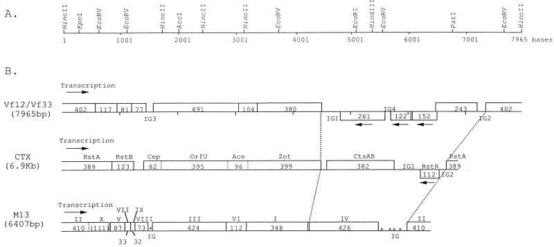
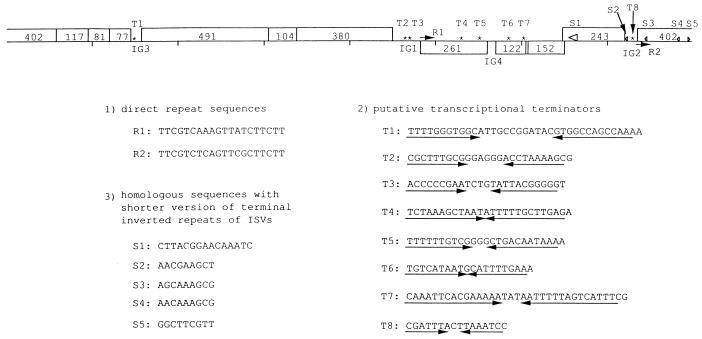
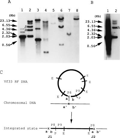
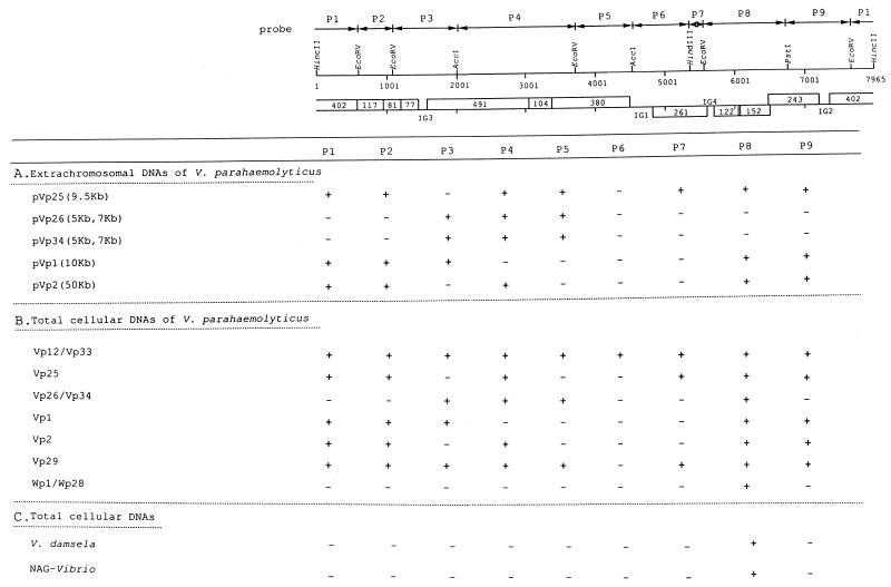
We have previously reported the isolation and characterization of two filamentous bacteriophages of Vibrio parahaemolyticus, designated Vf12 and Vf33. In this study, to understand the potential of these phages as tools for genetic transmission, we investigated the gene structures of replicative-form (RF) DNAs of their genomes and the distribution of these DNAs on chromosomal and extrachromosomal DNAs. The 7,965-bp nucleotide sequences of Vf12 and Vf33 were determined. An analysis of the overall gene structures revealed that Vf12 and Vf33 had conserved regions and distinctive regions. The gene organization of their conserved regions was similar to that of CTX phage of Vibrio cholerae and coliphage Ff of Escherichia coli, while their distinctive regions were characteristic of Vf12 and Vf33 phage genomes. Southern blot hybridization testing revealed that the filamentous phage genomes integrated into chromosomal DNA of V. parahaemolyticus at the distinctive region of the phage genome and were also distributed on some plasmids of V. parahaemolyticus and total cellular DNAs of one Vibrio damsela and one nonagglutinable Vibrio strain tested. These results strongly suggest the possibilities of genetic interaction among the bacteriophage Vf12 and Vf33 genomes and chromosomal and plasmid-borne DNAs of V. parahaemolyticus strains and of genetic transmission among strains through these filamentous phages.


Figures

FIG. 1
FIG. 1
Gene structures of bacteriophage Vf12 and Vf33 genomes. (A) Restriction enzyme cleavage map. The circular phage genome is represented in a linear form with the HincII site as point zero and is numbered in the 5′-to-3′ direction of the viral strand. (B) Comparison of the linear ORF maps of filamentous phages Vf12 and Vf33, CTX of V. cholerae, and M13 of E. coli. ORFs are represented as blocks. The numbers in the blocks refer to the number of predicted amino acids, and arrows indicate the transcription directions of genes. The genetic map of CTX phage was designed according to the work of Waldor and Mekalanos (34) and Waldor et al. (35). The genetic map of M13 phage was designed according to the work of Van Wezenbeek et al. (33).
FIG. 2
FIG. 2
Repeat sequences of the Vf12 and Vf33 genomes, which consist of (i) direct repeat sequences (R1, R2, and arrows), (ii) inverted repeat sequences (T1 to T8 and asterisks), and (iii) repeated sequences similar to the shorter versions of the 18-bp terminal inverted repeats of the ISVs (S1 to S5). S1 (large open arrowhead) is similar to a part of the inverted repeats (5′-GATTTACGCAACAAAGCC-3′). S2 to S5 indicate the homologous sequences with the shorter versions of the inverted repeats, which are indicated by ◃ (5′-AACAAAGCC-3′) and ▹ (5′-GGCTTTGTT-3′).
FIG. 3
FIG. 3
Hybridization patterns of the P8 (A) and P9 (B) probes and the mode of integration of the Vf12 and Vf33 genomes (C). (A and B) Vf12 and Vf33 phage genomes are present as the RF and concomitantly integrated into the chromosome. Numbers on the left are the sizes of λ HindIII markers. RF DNAs or total cellular DNAs digested with EcoRV were analyzed by agarose gel electrophoresis, transferred to nylon membranes, and hybridized with the labeled fragments P8 (A) and P9 (B). The chromosomal junction fragments of the Vp33 strain are labeled J1 and J2. (A) Lanes: 1, purified RF DNA of Vf33; 2, total cellular DNA of the Vp33 strain; 3, purified pVp25 DNA; 4, total cellular DNA of the Vp25 strain; 5, total cellular DNA of the Wp1 strain; 6, total cellular DNA of the Wp28 strain; 7, total cellular DNA of V. damsela; 8, total cellular DNA of NAG-Vibrio. (B) Lanes: 1, purified RF DNA of Vf33; 2, total cellular DNA of the Vp33 strain. The fragment labeled J1 has the same size as J1 of panel A, lane 2. (C) The phage genome is represented in a thick circular form, and that of the chromosome is in a thin linear form. P8 and P9 indicate the probes used in panels A and B, respectively. E and P indicate the EcoRV and PstI enzyme sites of the phage genome, respectively. a and b and a′ and b′ indicate the attachment sites of the phage genome and chromosome, respectively. J1 and J2 indicate the junction fragments that are the same as those of panels A and B.
FIG. 4
FIG. 4
Distribution of Vf12 and Vf33 genomes in extrachromosomal and chromosomal DNAs of V. parahaemolyticus and total cellular DNAs of other Vibrio species detected by Southern blot hybridization analysis. The cloned fragments used as probes (P1 to P9) are represented by solid lines with two arrowheads. + and − indicate the regions which hybridized and did not hybridize with probes, respectively. (A) Distribution on extrachromosomal DNAs of pVp1, pVp2, pVp25, pVp26, and pVp34. The numbers in parentheses refer to the sizes of the extrachromosomal DNAs. (B) Distribution on total cellular DNAs of V. parahaemolyticus. (C) Distribution on total cellular DNAs of V. damsela and NAG-Vibrio.

Similar articles

Cited by

References

    1. Baba K, Shirai H, Terai A, Kumagai K, Takeda Y, Nishibuchi M. Similarity of the tdh gene-bearing plasmids of Vibrio cholerae non-O1 and Vibrio parahaemolyticus. Microb Pathog. 1991;10:61–70. - PubMed
    1. Beck E, Zink B. Nucleotide sequence and genome organization of filamentous bacteriophages f1 and fd. Gene. 1981;16:35–58. - PubMed
    1. Birnboim H C, Doly J. A rapid alkaline extraction procedure for screening recombinant plasmid DNA. Nucleic Acids Res. 1979;7:1513–1523. - PMC - PubMed
    1. Blake P A, Weaver R E, Hollis D G. Diseases of humans (other than cholera) caused by vibrios. Annu Rev Microbiol. 1980;34:341–367. - PubMed
    1. Datz M, Janetzki-Mittmann C, Franke S, Gunzer F, Schmidt H, Karch H. Analysis of the enterohemorrhagic Escherichia coli O157 DNA region containing lambdoid phage gene p and Shiga-like toxin structural genes. Appl Environ Microbiol. 1996;62:791–797. - PMC - PubMed

Associated data

LinkOut - more resources