. 1998 Oct;180(19):5117-22.
doi: 10.1128/JB.180.19.5117-5122.1998.

Formation of single-stranded DNA during DNA transformation of Neisseria gonorrhoeae

Affiliations

Formation of single-stranded DNA during DNA transformation of Neisseria gonorrhoeae

M S Chaussee et al. J Bacteriol. 1998 Oct.

Abstract

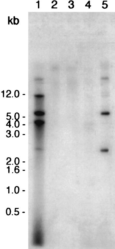
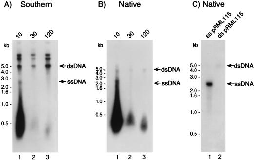
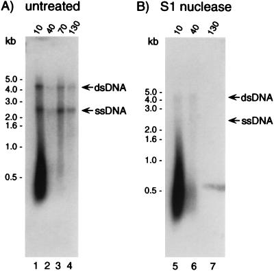
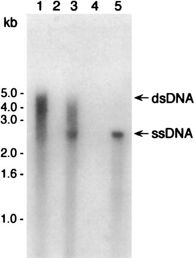
Neisseria gonorrhoeae is naturally competent for DNA transformation. In contrast to other natural prokaryotic DNA transformation systems, single-stranded donor DNA (ssDNA) has not previously been detected during transformation of N. gonorrhoeae. We have reassessed the physical nature of gonococcal transforming DNA by using a sensitive nondenaturing native blotting technique that detects ssDNA. Consistent with previous analyses, we found that the majority of donor DNA remained in the double-stranded form, and only plasmid DNAs that carried the genus-specific DNA uptake sequence were sequestered in a DNase I-resistant state. However, when the DNA was examined under native conditions, S1 nuclease-sensitive ssDNA was identified in all strains tested except for those bacteria that carried the dud-1 mutation. Surprisingly, ssDNA was also found during transformation of N. gonorrhoeae comA mutants, which suggested that ssDNA was initially formed within the periplasm.


Figures

FIG. 1
FIG. 1
DUS-specific uptake of DNA into a DNase I-resistant state by competent gonococci. N. gonorrhoeae wild-type and N. gonorrhoeae dud-1 strains were resuspended in transformation medium that contained pRML115 (DUS+) or, as negative controls, pRML110 (DUS) or no transforming DNA for 20 min at 37°C. Following addition of DNase I to degrade extracellular DNA, DNase I-resistant DNA was isolated and analyzed by Southern blotting with a probe specific to the ermC gene present in both pRML115 and pRML110. Lanes: 1, MS11 incubated with pRML115 (DUS+); 2, MS11 incubated with pRML110 (DUS); 3, MS11 (no transforming DNA added); 4, MS11 dud-1 incubated with pRML115 (DUS+); 5, MS11 lysate plus pRML115 (positive control). The blot was exposed to film at −70°C for approximately 20 h.
FIG. 2
FIG. 2
Southern blot and native blot analyses of DNase I-resistant DNA. MS11 was incubated with pRML115 (DUS+) for 10 min at 37°C. DNase I was added, samples were removed 10, 30, and 120 min after the addition of DNase I, and total nucleic acid was isolated. The DNase I-resistant DNA was subsequently analyzed by Southern blotting (A) and native blotting (B) with an ermC-specific radiolabeled probe. (C) As a control, native blotting of 5 ng of linear alkali-denatured pRML115 reacted with the ermC probe (lane 1), in contrast to 5 ng of linear double-stranded pRML115 that did not react with probe (lane 2). The arrows to the right of each panel indicate the positions of migration of both double- and single-stranded pRML115. The Southern blot and native blot were exposed to film at −70°C for approximately 18 and 36 h, respectively.
FIG. 3
FIG. 3
S1 nuclease treatment of DNase I-resistant DNA. Following transformation of strain MS11 with pRML115, samples were removed 10, 40, 70, and 130 min after the addition of DNase I. Selected DNA samples were treated with S1 nuclease prior to electrophoresis and were compared to untreated samples under native conditions with an ermC-specific probe. (A) Untreated DNA; (B) S1 nuclease-treated DNA. The arrows to the right of each panel indicate the positions of migration of both double- and single-stranded pRML115. The blots were exposed to film overnight at −70°C.
FIG. 4
FIG. 4
ssDNA is formed by comA mutants of gonococci. Following the incubation of P9 and P9 comA with pRML115 (DUS+), DNase I-resistant DNA was analyzed by native blotting with an ermC-specific probe. Lane 1, P9; lane 2, P9 treated with S1 nuclease; lane 3, P9 comA; lane 4, P9 comA treated with S1 nuclease; lane 5, alkali-denatured pRML115 (control). The blot was exposed to film at −70°C for approximately 96 h.

References

    1. Biswas G D, Burnstein K L, Sparling P F. Linearization of donor DNA during plasmid transformation in Neisseria gonorrhoeae. J Bacteriol. 1986;168:756–761. - PMC - PubMed
    1. Biswas G D, Sox T, Blackman E, Sparling P F. Factors affecting genetic transformation of Neisseria gonorrhoeae. J Bacteriol. 1977;129:983–992. - PMC - PubMed
    1. Biswas G D, Sparling P F. Entry of double-stranded deoxyribonucleic acid during transformation of Neisseria gonorrhoeae. J Bacteriol. 1981;145:638–640. - PMC - PubMed
    1. Butler C A, Gotschlich E C. High-frequency mobilization of broad-host-range plasmids into Neisseria gonorrhoeae requires methylation in the donor. J Bacteriol. 1991;173:5793–5799. - PMC - PubMed
    1. Danner D B, Deich R A, Sisco K L, Smith H O. An eleven-base-pair sequence determines the specificity of DNA uptake in Haemophilus transformation. Gene. 1980;11:311–318. - PubMed

MeSH terms

LinkOut - more resources