. 1998 Sep 29;95(20):11566-71.
doi: 10.1073/pnas.95.20.11566.

Proteolytic activation of PKN by caspase-3 or related protease during apoptosis

Affiliations

Proteolytic activation of PKN by caspase-3 or related protease during apoptosis

M Takahashi et al. Proc Natl Acad Sci U S A. .

Abstract

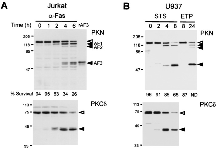
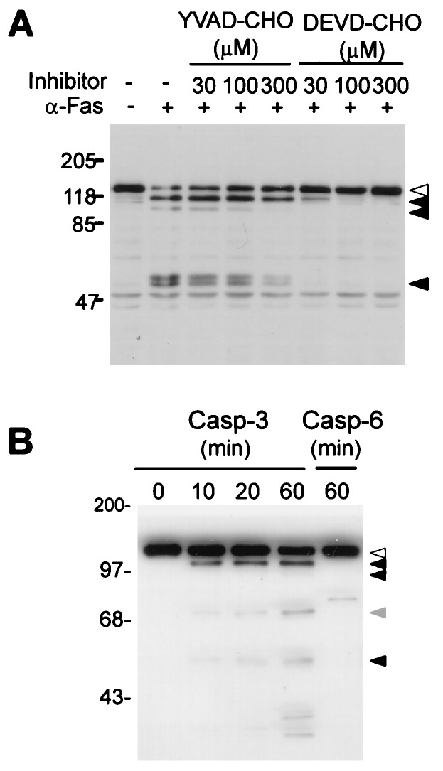
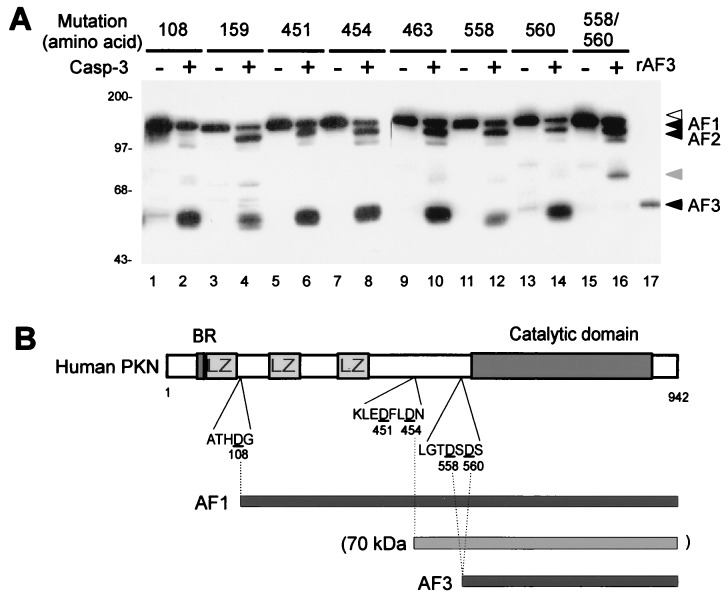
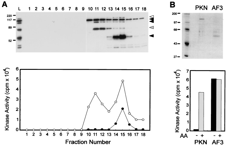
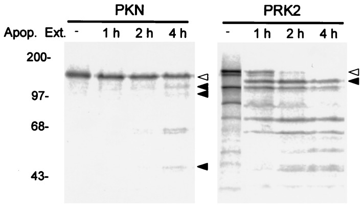
PKN, a fatty acid- and Rho-activated serine/threonine kinase having a catalytic domain highly homologous to protein kinase C (PKC), was cleaved at specific sites in apoptotic Jurkat and U937 cells on Fas ligation and treatment with staurosporin or etoposide, respectively. The cleavage of PKN occurred with a time course similar to that of PKCdelta, a known caspase substrate. This proteolysis was inhibited by a caspase inhibitor, acetyl-Asp-Glu-Val-Asp-aldehyde. The cleavage fragments were generated in vitro from PKN by treatment with recombinant caspase-3. Site-directed mutagenesis of specific aspartate residues prevented the appearance of these fragments. These results indicate that PKN is cleaved by caspase-3 or related protease during apoptosis. The major proteolysis took place between the amino-terminal regulatory domain and the carboxyl-terminal catalytic domain, and it generated a constitutively active kinase fragment. The cleavage of PKN may contribute to signal transduction, eventually leading to apoptosis.


Figures

Figure 1
Figure 1
Proteolytic cleavage of PKN in apoptotic cells. (A) Cleavage of PKN in Jurkat cells. Jurkat cells were treated with 150 ng/ml α-Fas mAb for the indicated time periods. (B) Cleavage of PKN in U937 cells. U937 cells were treated with 1 μM staurosporin (STS) or 10 μg/ml etoposide (ETP) for the indicated time periods. Cell lysates were subjected to immunoblot analysis with αC6 (Upper) or nPKCδ (C-20) (Lower). White and black arrowheads on the right indicate uncleaved and cleaved proteins, respectively. The cleavage products of PKN designated as AF1, AF2, and AF3 are shown with the black arrowheads in A. rAF3 indicates the recombinant AF3 expressed in COS-7 cells. Percentage of the viable cells determined by Annexin V-FITC apoptosis kit at each timepoint is given below each lane of PKN blots. ND, not determined. Molecular mass markers in kDa are indicated on the left of blots.
Figure 2
Figure 2
Cleavage of PKN by caspase-3-like protease. (A) Inhibition of PKN cleavage during apoptosis by DEVD-CHO. Jurkat cells were pretreated with various concentrations of the tetrapeptide inhibitor of caspase-1 (YVAD-CHO) or caspase-3 (DEVD-CHO) for 1 h, as indicated, and then incubated with α-Fas mAb (150 ng/ml) for an additional 6 h. The cleavage of PKN was analyzed by immunoblotting with αC6. (B) In vitro cleavage of PKN by recombinant caspases. His-PKN (40 ng) was incubated with recombinant His-caspase-3 (Casp-3) or GST-caspase-6 (Casp-6) at 30°C for the indicated time periods, then analyzed by immunoblotting with PKN (C-19). The cleavage products corresponding to AF1, AF2, and AF3 were observed from 10-min incubation with caspase-3, while a cleavage product of a different size was observed from caspase-6 cleavage. White and black arrowheads on the right of the blot indicate the proteins uncleaved and cleaved by caspase-3, respectively. Gray arrowhead indicates a caspase-3 cleavage product that was not detected in vivo. Molecular mass markers in kDa are indicated on the left.
Figure 3
Figure 3
Determination of the caspase-3 cleavage sites in PKN. (A) Cleavage of PKN point mutants replacing Asp with Ala by caspase-3. Extracts of COS-7 cells expressing the FLAG-tagged PKN mutants were incubated with or without recombinant His-caspase-3 (Casp-3), and were analyzed for the cleavage of PKN by immunoblotting with PKN (C-19). Mutated amino acid number is shown on the top. White and black arrowheads on the right of the blot indicate the uncleaved and cleaved bands, respectively. Gray arrowhead indicates the cleavage product detected only in the in vitro reaction. rAF3 indicates the recombinant AF3-FLAG expressed in COS-7 cells. Molecular mass markers in kDa are indicated on the left. (B) Schematic representation of the structure of human PKN with the predicted cleavage sites and locations of AF1, AF3, and the in vitro 70-kDa product. The mutated Asp residues giving rise to the resistance to the cleavage are presented with neighboring amino acid sequences below the structure of PKN. The predicted positions of AF1, AF3, and the 70-kDa fragment are shown on the bottom. LZ, leucine zipper-like motif; BR, basic region.
Figure 4
Figure 4
Generation of constitutively active kinase after caspase-3-mediated proteolysis of PKN. (A) Mono Q column chromatography of the in vitro cleavage products of PKN. Baculovirus-expressed His-GST-PKN was digested with His-caspase-3, and the cleavage products were separated by Mono Q column chromatography. Each fraction was subjected to immunoblotting with αC6 (Upper) and the kinase assay in the presence (○) or absence (•) of AA (Lower). L indicates the sample loaded to the column. Positions of uncleaved and cleaved proteins are indicated with black and white arrowheads, respectively. Gray arrowhead indicates a cleavage product detected only in the in vitro reaction. (B) comparison of kinase activity between the recombinant full length PKN and its mutant AF3. PKN-FLAG (PKN) and AF3-FLAG (AF3) expressed in COS-7 cells were purified by anti-FLAG column, then equal amount of the proteins were subjected to SDS/PAGE followed by Coomassie brilliant blue staining (Upper) and the kinase assay in the presence or absence of AA (Lower). The results are representative of three independent experiments. Molecular mass markers in kDa are indicated on the left of the gel.
Figure 5
Figure 5
Cleavage of PKN and PRK2 by apoptotic extracts from Jurkat cells treated with α-Fas mAb. In vitro translated PKN and PRK2 were incubated with apoptotic extracts (Apop. Ext.) prepared from Jurkat cells treated with 150 ng/ml α-Fas mAb for the indicated time periods. White and black arrowheads indicate uncleaved and cleaved proteins, respectively. Molecular mass markers in kDa are indicated on the left.

References

    1. Mukai H, Ono Y. Biochem Biophys Res Commun. 1994;199:897–904. - PubMed
    1. Mukai H, Kitagawa M, Shibata H, Takanaga H, Mori K, Shimakawa M, Miyahara M, Hirao K, Ono Y. Biochem Biophys Res Commun. 1994;204:348–356. - PubMed
    1. Kitagawa M, Mukai H, Shibata H, Ono Y. Biochem J. 1995;310:657–664. - PMC - PubMed
    1. Mukai H, Mori K, Takanaga H, Kitagawa M, Shibata H, Shimakawa M, Miyahara M, Ono Y. Biochim Biophys Acta. 1995;1261:296–300. - PubMed
    1. Palmer R H, Ridden J, Parker P J. Eur J Biochem. 1995;227:344–351. - PubMed

Publication types

MeSH terms