. 1998 Sep 29;95(20):11607-12.
doi: 10.1073/pnas.95.20.11607.

ATP hydrolysis catalyzed by human replication factor C requires participation of multiple subunits

Affiliations

ATP hydrolysis catalyzed by human replication factor C requires participation of multiple subunits

J Cai et al. Proc Natl Acad Sci U S A. .

Abstract

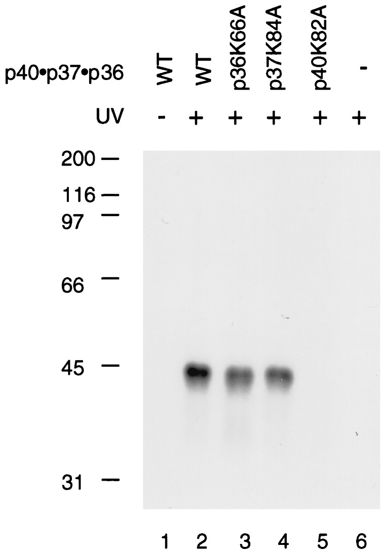
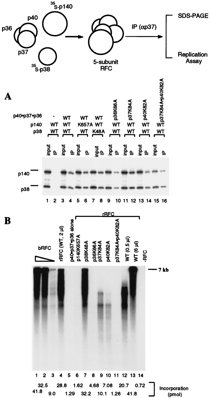
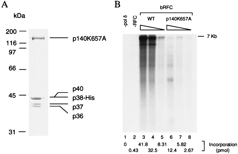
Human replication factor C (hRFC) is a five-subunit protein complex (p140, p40, p38, p37, and p36) that acts to catalytically load proliferating cell nuclear antigen onto DNA, where it recruits DNA polymerase delta or epsilon to the primer terminus at the expense of ATP, leading to processive DNA synthesis. We have previously shown that a subcomplex of hRFC consisting of three subunits (p40, p37, and p36) contained DNA-dependent ATPase activity. However, it is not clear which subunit(s) hydrolyzes ATP, as all five subunits include potential ATP binding sites. In this report, we introduced point mutations in the putative ATP-binding sequences of each hRFC subunit and examined the properties of the resulting mutant hRFC complex and the ATPase activity of the hRFC or the p40.p37.p36 complex. A mutation in any one of the ATP binding sites of the p36, p37, p40, or p140 subunits markedly reduced replication activity of the hRFC complex and the ATPase activity of the hRFC or the p40.p37.p36 complex. A mutation in the ATP binding site of the p38 subunit did not alter the replication activity of hRFC. These findings indicate that the replication activity of hRFC is dependent on efficient ATP hydrolysis contributed to by the action of four hRFC subunits.


Figures

Figure 1
Figure 1
(A) Mutagenesis in the Walker A motifs of the p36, p37, and p40 subunits. The amino acid sequences of the Walker A motifs in the p36, p37, and p40 subunits are aligned. The conserved lysine residue (boxed) in each subunit was changed to alanine by site-directed mutagenesis. (B) Purification of wt and mutant p40·p37·p36 complexes. The purified wt and mutant p40·p37·p36 complexes (described in Materials and Methods) were analyzed by SDS/9% polyacrylamide gels. Lane 1, 3 μg of wt p40·p37·p36 complex; lanes 2–4, 3 μg of the mutant p40·p37·p36 complex with a mutation in p36 (p36K66A), p37 (p37K84A), and p40 (p40K82A), respectively. (C) ATPase activity of wt and mutant p40·p37·p36 complexes. ATPase measurements were carried out as described in Materials and Methods. Reaction mixtures contained wt and mutant p40·p37·p36 complexes in amounts as indicated, in the presence of 12.5 μM (as nucleotides) φX174 ssc DNA.
Figure 2
Figure 2
ATP binds to the p40 subunit in the p40·p37·p36 complex. Reactions containing [α-32P]ATP and the wt or various mutant p40·p37·p36 complexes (0.3 μg) were UV-irradiated and subjected to SDS-PAGE as described in the Materials and Methods. Lane 1, wt p40·p37·p36 complex not UV-irradiated; lane 2, wt p40·p37·p36; lane 3, p36K66A; lane 4, p37K84A; lane 5, p40K82A; lane 6, no protein added.
Figure 3
Figure 3
Coupled in vitro transcription-translation reactions with plasmid DNA containing the coding sequence for the hRFC p140 and p38 subunits were carried out in the presence or absence of the p40·p37·p36 complex followed by immunoprecipitation using a polyclonal antibody against the hRFC p37 subunit. Because the C-terminal half of the p140 subunit (amino acids 555-1147) forms an hRFC complex that is 5–10 fold more active than that formed with the full-length p140 subunit, p140N555 was used in the experiments described here. The immunoprecipitated products were analyzed on SDS/9% polyacrylamide gels followed by autoradiography to visualize the 35S-labeled p140 and p38, or assayed for their replication activities. (A) Reconstitution of hRFC from its five subunits. Reactions containing 20% of the input material (lanes 1, 3, 5, 7. 9, 11, 13, and 15) and the immunoprecipitates (lanes 2, 4, 6, 8, 10, 12, 14, and 16) were separated by SDS/9% polyacrylamide gels. The reactions contained wt or mutant hRFC subunits or subcomplexes as indicated. (B) Replication activities of mutant hRFCs. Mutant hRFCs, immunoprecipitated on protein A beads, were examined for their ability to elongate singly primed M13 DNA as described in Materials and Methods. Reactions shown in lanes 1–3 were carried out in the presence 70, 14, and 1.4 fmol of baculovirus RFC, respectively. Reactions shown in lanes 4–11 were carried out after immunoprecipitation of 2 μl of the reconstitution mixture as follows: lane 4, RFC reconstituted with wt p140, p38, and wt p40·p37·p36 complex; lane 5, the wt p40·p37·p36 complex only; lane 6, mutant p140 (p140K657A), wt p38, and wt p40·p37·p36 complex; lane 7, wt p140, mutant p38 (p38K48A), and wt p40·p37·p36 complex; lane 8, wt p140 and p38, mutant p40·p37·p36 complex (p36K66a); lane 9, wt p140 and p38, mutant p40·p37·p36 complex (p37K84A); lane 10, wt p140 and p38, mutant p40·p37·p36 complex (p40K82A); lane 11, wt p140 and p38, mutant p40·p37·p36 complex (p37K84Ap40K82A). Reactions shown in lanes 12 and 13 were carried out after immunoprecipitation of two levels of the reconstituted mixture (0.5 and 6 μl, respectively) containing wt p140, p38, and wt p40·p37·p36 complex. The reaction shown in lane 14 was carried out in the absence of RFC. Total nucleotide incorporation (pmol) obtained in the above reactions, detected after acid precipitation and liquid scintillation counting, are shown at the bottom of the figure.
Figure 4
Figure 4
(A) Purification of hRFCp140K657A. Mutant hRFC was purified from baculovirus-infected high five insect cells cells as described in the Materials and Methods. The purified product (2 μg) was separated by SDS/9% polyacrylamide gels followed by Coomasie staining. (B) Replication activity of mutant hRFC. The wt and mutant hRFCs were examined for their ability to support DNA synthesis from singly primed M13 DNA as described in Materials and Methods. Lanes 1 and 2 represent reactions carried out in the absence of pol δ or RFC, respectively. Wild-type baculovirus RFC was added to the reactions in amount as follows: lane 3, 70 fmol; lane 4, 14 fmol; lane 5, 1.4 fmol. Mutant baculovirus RFC was added to the reactions in amounts as follows: lane 6, 333 fmol; lane 7, 67 fmol; lane 8, 23 fmol. Total nucleotide incorporation (pmol), detected after acid precipitation and liquid scintillation counting, is shown at the bottom of the figure.
Figure 5
Figure 5
A model for the loading of PCNA onto DNA catalyzed by RFC. The RFC subunits that interact with PCNA are indicated in the figure (A and B). Class A subunits can interact with PCNA in the absence of ATP. ATP binding induces a conformational change in RFC that permit the interaction of class B subunits with PCNA. The PCNA ring opens and recloses in response to interactions with the class B subunits governed by ATP binding and hydrolysis. Multiple rounds of ATP binding and hydrolysis may be required before PCNA is finally loaded onto DNA.

References

    1. Lee S-H, Eki T, Hurwitz J. Proc Natl Acad Sci USA. 1989;86:7361–7365. - PMC - PubMed
    1. Tsurimoto T, Stillman B. Mol Cell Biol. 1989;9:609–619. - PMC - PubMed
    1. Lee S-H, Kwong A D, Pan Z-Q, Hurwitz J. J Biol Chem. 1991;266:584–602. - PubMed
    1. Tsurimoto T, Stillman B. Proc Natl Acad Sci USA. 1990;87:1023–1027. - PMC - PubMed
    1. Yoder B L, Burgers P M. J Biol Chem. 1991;266:22689–22697. - PubMed

Publication types

MeSH terms

LinkOut - more resources