Comparative Study
. 1998 Sep 29;95(20):11637-42.
doi: 10.1073/pnas.95.20.11637.

The endoribonucleolytic N-terminal half of Escherichia coli RNase E is evolutionarily conserved in Synechocystis sp. and other bacteria but not the C-terminal half, which is sufficient for degradosome assembly

Affiliations
Comparative Study

The endoribonucleolytic N-terminal half of Escherichia coli RNase E is evolutionarily conserved in Synechocystis sp. and other bacteria but not the C-terminal half, which is sufficient for degradosome assembly

V R Kaberdin et al. Proc Natl Acad Sci U S A. .

Abstract

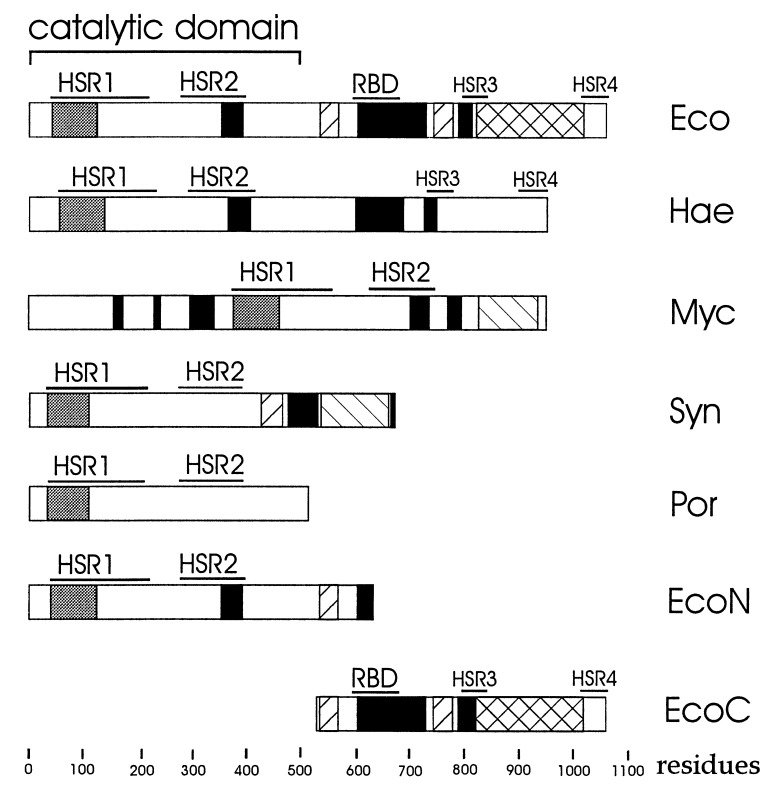
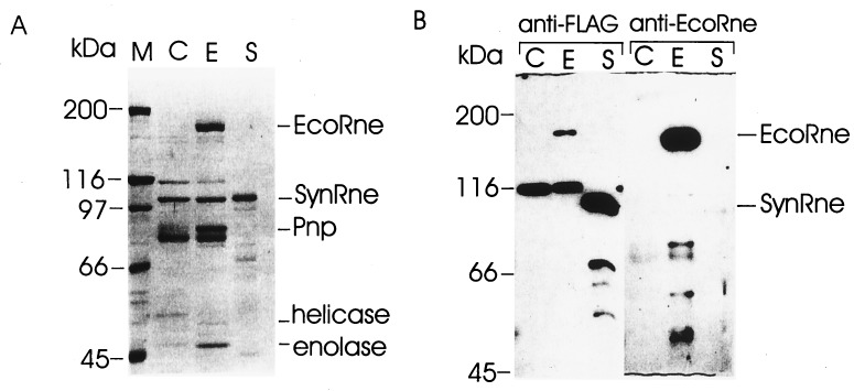
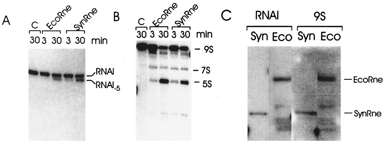
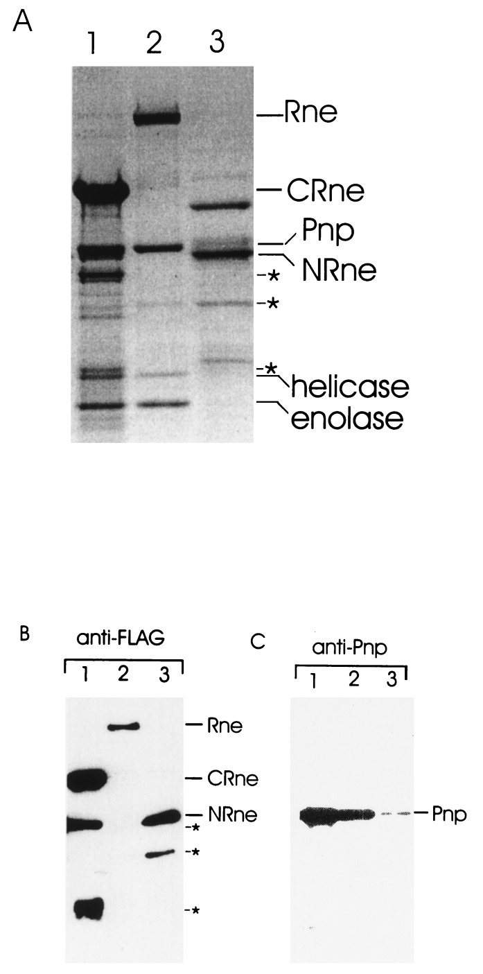
Escherichia coli RNase E, an essential single-stranded specific endoribonuclease, is required for both ribosomal RNA processing and the rapid degradation of mRNA. The availability of the complete sequences of a number of bacterial genomes prompted us to assess the evolutionarily conservation of bacterial RNase E. We show here that the sequence of the N-terminal endoribonucleolytic domain of RNase E is evolutionarily conserved in Synechocystis sp. and other bacteria. Furthermore, we demonstrate that the Synechocystis sp. homologue binds RNase E substrates and cleaves them at the same position as the E. coli enzyme. Taken together these results suggest that RNase E-mediated mechanisms of RNA decay are not confined to E. coli and its close relatives. We also show that the C-terminal half of E. coli RNase E is both sufficient and necessary for its physical interaction with the 3'-5' exoribonuclease polynucleotide phosphorylase, the RhlB helicase, and the glycolytic enzyme enolase, which are components of a "degradosome" complex. Interestingly, however, the sequence of the C-terminal half of E. coli RNase E is not highly conserved evolutionarily, suggesting diversity of RNase E interactions with other RNA decay components in different organisms. This notion is supported by our finding that the Synechocystis sp. RNase E homologue does not function as a platform for assembly of E. coli degradosome components.


Figures

Figure 1
Figure 1
Structure of E. coli Rne (Eco) and proteins having similar sequences in Haemophilus influenzae Rd (Hae), Mycobacterium tuberculosis (Myc), Synechocystis sp. (Syn), Porphyra purpurea (Por). The N- and C-terminal segments of E. coli RNase E (EcoN and EcoC, respectively) also are shown. Horizontal bars indicate the location of two regions (designated HSR1 and HSR2; see Table 1) within the N-terminal endoribonucleolytic domain of E. coli RNase E, which are highly conserved in other bacteria, two regions (designated HSR3 and HSR4) within the C-terminal half of E. coli RNase E having significant sequence similarity to the corresponding region of the H. influenzae Rd Rne homologue, and an RNA-binding domain of E. coli RNase E (16). The putative S1-like RNA-binding domain predicted by Bycroft et al. (20) and regions enriched in arginine, proline, and acidic residues (see Table 1) are indicated by gray, black, and back-hatched and forward-hatched boxes, respectively.
Figure 2
Figure 2
Purification of the Synechocystis sp. RNase E. E. coli cell extracts lacking overexpressed protein (C) or containing overexpressed E. coli (E) or Synechocystis sp. (S) RNase E were treated with micrococcal nuclease and purified on the anti-FLAG gel column as described (12). The preparations were run in triplicate in a 8% polyacrylamide/SDS gel. One-third of the gel was stained with Coomassie brilliant blue (A), whereas the remainder of the gel was used for blotting the proteins onto poly(vinylidene difluoride) membrane, and the remaining two-thirds probed either with anti-FLAG or anti-EcoRne antibodies (B). Indicated are protein size markers (kDa), Synechocystis sp. Rne (SynRne), and the major components of the E. coli degradosome; RNase E (EcoRne), polynucleotide phosphorylase (Pnp), the RhlB RNA helicase, and enolase. Although SynRne migrates in SDS/polyacrylamide gels with an apparent molecular mass of 105 kDa, the predicted molecular mass of SynRne is 78 kDa. A possible explanation for this observation is that SynRne contains a region rich in prolines, which have been shown to affect the migration of other proteins, including E. coli RNase E (22).
Figure 3
Figure 3
In vitro cleavage of RNAI (A) and 9S RNA (B) by the E. coli and Synechocystis sp. Rne proteins. Internally labeled RNAI or 9S RNA was incubated in RNase E reaction buffer for 3 and 30 min with either E. coli RNase E (EcoRne) or Synechocystis sp. Rne (SynRne), or without protein for 30 min (C), as a negative control. The positions of the original substrates (RNAI and 9S RNA) and their cleavage products (RNA I-5 and 5S rRNA (5S), respectively) are indicated. (C) Protein blots probed with RNAI and 9S RNA. Samples of the Synechocystis sp. Rne protein (Syn) and the E. coli RNA degradosome (Eco) were resolved by SDS/PAGE, electroblotted onto poly(vinylidene difluoride) membrane, and probed with 32P-labeled RNAI or 9S RNA as described in Materials and Methods. The Synechocystis sp. Rne (SynRne) and E. coli Rne (EcoRne) proteins complexed with RNA are indicated.
Figure 4
Figure 4
Identification of the proteins associated with the C-terminal half of E. coli Rne. Cell extracts containing overexpressed full-length E. coli Rne or N- or C-terminal portions of this polypeptide (see Fig. 1) were treated with micrococcal nuclease and purified by affinity chromatography as described in Materials and Methods. Samples of each preparation were run in triplicate on an 8% polyacrylamide/SDS gel (lanes 2, 3, and 1, respectively). As described for Fig. 2B, one-third of the gel was stained with Coomassie brilliant blue (A), whereas the remaining two-thirds were either probed with anti-FLAG (B) or anti-Pnp (C) antibodies after transfer to a poly(vinylidene difluoride) membrane. The position of the major components of the E. coli RNA degradosome, RNase E (Rne), polynucleotide phosphorylase (Pnp), enolase, RhlB helicase as well as N- and C-terminal fragments (NRne and CRne, respectively) of E. coli Rne are indicated. The proteins marked by asterisks and detected by Western blot with anti-FLAG antibodies are proteolytic products of NRne and CRne.

References

    1. Cohen S N, McDowall K J. Mol Microbiol. 1997;23:1099–1106. - PubMed
    1. Apirion D, Lassar A B. J Biol Chem. 1978;253:1738–1742. - PubMed
    1. Ehretsmann C P, Carpousis A J, Krisch H M. FASEB J. 1992;6:3186–3192. - PubMed
    1. Melefors Ö, Lundberg U, von Gabain A. In: Control of Messenger RNA Stability. Belasco J, Brawerman G, editors. San Diego: Academic; 1993. pp. 53–70.
    1. Lin-Chao S, Cohen S N. Cell. 1991;65:1233–1242. - PubMed

Publication types

LinkOut - more resources