. 1998 Sep 29;95(20):11673-8.
doi: 10.1073/pnas.95.20.11673.

Lipid patches in membrane protein oligomers: crystal structure of the bacteriorhodopsin-lipid complex

Affiliations

Lipid patches in membrane protein oligomers: crystal structure of the bacteriorhodopsin-lipid complex

L Essen et al. Proc Natl Acad Sci U S A. .

Abstract

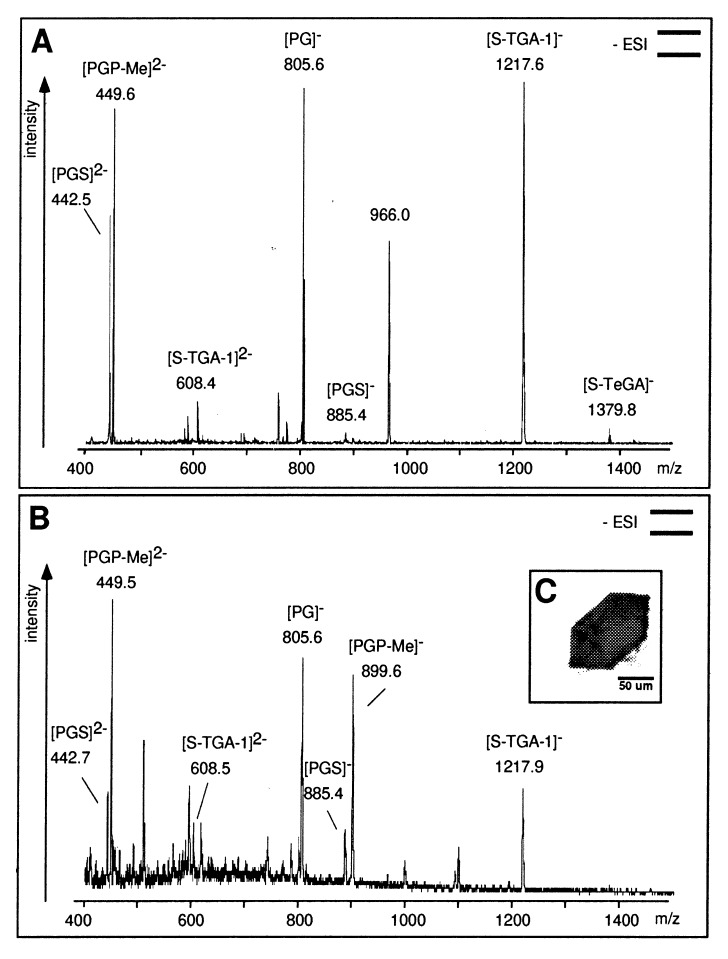
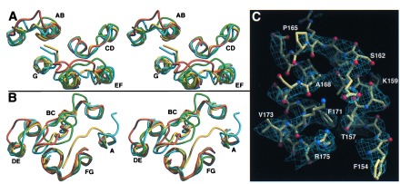
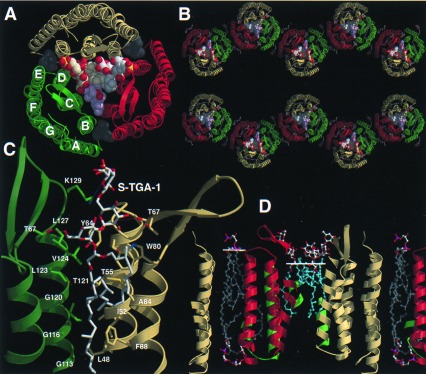
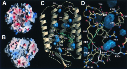
Heterogenous nucleation on small molecule crystals causes a monoclinic crystal form of bacteriorhodopsin (BR) in which trimers of this membrane protein pack differently than in native purple membranes. Analysis of single crystals by nano-electrospray ionization-mass spectrometry demonstrated a preservation of the purple membrane lipid composition in these BR crystals. The 2.9-A x-ray structure shows a lipid-mediated stabilization of BR trimers where the glycolipid S-TGA-1 binds into the central compartment of BR trimers. The BR trimer/lipid complex provides an example of local membrane thinning as the lipid head-group boundary of the central lipid patch is shifted by 5 A toward the membrane center. Nonbiased electron density maps reveal structural differences to previously reported BR structures, especially for the cytosolic EF loop and the proton exit pathway. The terminal proton release complex now comprises an E194-E204 dyad as a diffuse proton buffer.


Figures

Figure 1
Figure 1
(A) Negative-ion nanoESI-MS of lipid extracts from PM. The phospholipids PGS and PGP-Me show signals at m/z 442.5, 449.5 (z = 2), and 885.6. The signal at m/z 899.5 is absent because of the double-negative state of detected PGP-Me. PG, the major hydrolysis product of PGS and PGP-Me, was detected at m/z 805.6; phosphatidic acid (PA, m/z 731.5) is absent. The sulfated glycolipids S-TGA-1 and S-TeGA (sulfated tetraglycosyldiphytanylglycerol, a minor species in PM) show signals at m/z 1217.6 and 1379.8. The phospho- and glycolipids were identified by class-specific fragmentations in tandem MS (MS/MS) analyses at their optimized collision energies. (B) NanoESI-MS of the lipid extract of the BR crystal used for crystallographic data collection (shown in C). Pictograms summarize the experimental setup according to ref. .
Figure 2
Figure 2
Comparison between monomer B of the BR trimer (yellow) and recent BR structures (green, ref. ; cyan, ref. ; orange, ref. 23). The stereodiagrams show the cytosolic (A) and extracellular (B) surface loops. (C) Conformation of the EF loop and the cytosolic ends of helices E and F. The sigmaa-weighted 2Fobs-Fcalc electron density (14) is contoured at 0.175 e/Å3. Figs. 2–4 were made with molscript (42) and raster3d (43).
Figure 3
Figure 3
Lipid binding and layer packing of BR trimers in PM and monoclinic BR crystals. (A) Top view on the BR trimer/lipid complex from the extracellular side. Lipids are shown as space-filling models. Single phytanols (gray) are located on the cytosolic side of the BR trimer. (B) View along the a*-axis of the monoclinic BR crystal (monomer A, yellow; B, green; C, red). (C) Binding site of the glycolipid S-TGA-1 as viewed from the trimer axis. (D) Cross section of the PM model. Monomer B and an associated S-TGA-1 lipid replaced BR and the lipids 261, 266 of the previous PM model of Grigorieff et al. (11). The white lines show the head group/lipid boundaries.
Figure 4
Figure 4
Proton conductance in the BR trimer. (A) Proton collection surface of the cytosolic side. (B) Proton exit surface of the extracellular side. Electrostatic surface potentials were calculated with grasp (44) and shown from −20 kcal/mol (red) to +20 kcal/mol (blue). The internal residues D96, D115, and E204 were assumed to be protonated. (C) Internal cavities of BR. Twelve cavities were found by voidoo (45) using a probe radius of 1 Å and a primary grid spacing of 0.25 Å for enhanced sensitivity. Eight cavities (blue) fulfilled the criteria to be discontinuous from the molecular surface and capable of accommodating at least one water. White numbers indicate the maximal number of waters per cavity. (D) Proton exit pathway with the E194/E204 dyad (white). Cavities are shown in blue, H bonds within a 3.5-Å cutoff in white.

References

    1. Oesterhelt D, Stoeckenius W. Proc Natl Acad Sci USA. 1973;70:2853–2857. - PMC - PubMed
    1. Oesterhelt D, Tittor J, Bamberg E. J Bioenerg Biomemb. 1992;24:181–191. - PubMed
    1. Lanyi J K. J Biol Chem. 1997;272:31209–31212. - PubMed
    1. Oesterhelt D, Bräuchle C, Hampp N. Q Rev Biophys. 1991;24:425–478. - PubMed
    1. Hartmann R, Sickinger H-D, Oesterhelt D. FEBS Lett. 1977;82:1–6. - PubMed

LinkOut - more resources