. 1998 Sep 29;95(20):11679-84.
doi: 10.1073/pnas.95.20.11679.

Conformational gating of the electron transfer reaction QA-.QB --> QAQB-. in bacterial reaction centers of Rhodobacter sphaeroides determined by a driving force assay

Affiliations

Conformational gating of the electron transfer reaction QA-.QB --> QAQB-. in bacterial reaction centers of Rhodobacter sphaeroides determined by a driving force assay

M S Graige et al. Proc Natl Acad Sci U S A. .

Abstract

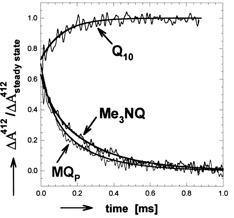
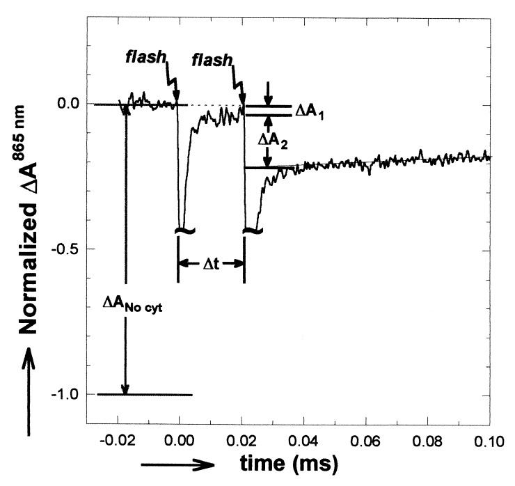
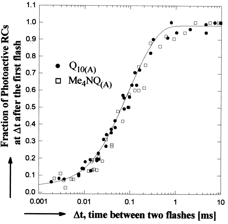
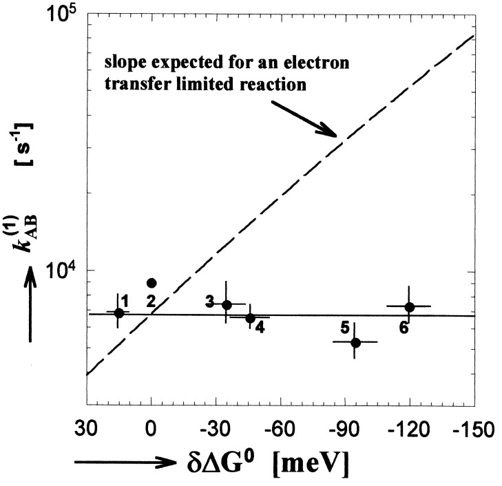
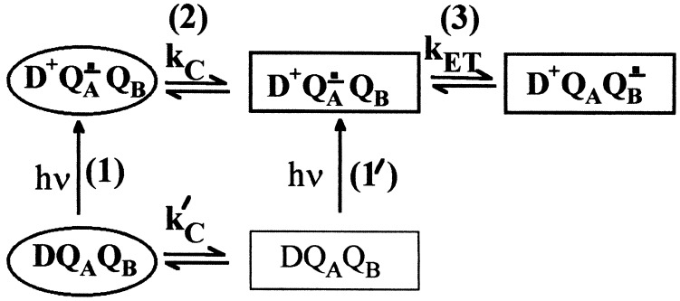
The mechanism of the electron transfer reaction, QA-.QB --> QAQB-., was studied in isolated reaction centers from the photosynthetic bacterium Rhodobacter sphaeroides by replacing the native Q10 in the QA binding site with quinones having different redox potentials. These substitutions are expected to change the intrinsic electron transfer rate by changing the redox free energy (i.e., driving force) for electron transfer without affecting other events that may be associated with the electron transfer (e.g., protein dynamics or protonation). The electron transfer from QA-. to QB was measured by three independent methods: a functional assay involving cytochrome c2 to measure the rate of QA-. oxidation, optical kinetic spectroscopy to measure changes in semiquinone absorption, and kinetic near-IR spectroscopy to measure electrochromic shifts that occur in response to electron transfer. The results show that the rate of the observed electron transfer from QA-. to QB does not change as the redox free energy for electron transfer is varied over a range of 150 meV. The strong temperature dependence of the observed rate rules out the possibility that the reaction is activationless. We conclude, therefore, that the independence of the observed rate on the driving force for electron transfer is due to conformational gating, that is, the rate limiting step is a conformational change required before electron transfer. This change is proposed to be the movement, controlled kinetically either by protein dynamics or intermolecular interactions, of QB by approximately 5 A as observed in the x-ray studies of Stowell et al. [Stowell, M. H. B., McPhillips, T. M., Rees, D. C., Soltis, S. M., Abresch, E. & Feher, G. (1997) Science 276, 812-816].


Figures

Figure 1
Figure 1
Optical absorbance changes at 412 nm associated with electron transfer from QA to QB in hybrid (Me3NQ or MQP in the QA site and Q10 in the QB site) and native (Q10 in both QA and QB sites) RCs. In native RCs, the absorbance change is due to an electrochromic shift of the BPhe absorption. In hybrid RCs, the main effect is caused by the difference in extinction coefficient between the naphthosemiquinones (NQ in the QA site and Q10 in the QB site). The observed transients in hybrid RCs are reduced by the electrochromic shift, which was assumed to be the same as that observed in native RCs. The data for the hybrid RCs were normalized to the expected absorption determined from the known difference in extinction coefficients between (NQ)A and Q10 (11, 20, 21). A shallow slope determined by monitoring the recombination reactions (D+QA → DQA and D+QAQB → DQAQB) was subtracted for clarity. Addition of terbutryne, which inhibits electron transfer to QB, eliminated the transient kinetic phases. [Conditions: 5 μM 2.4.1RCs, BMK buffer (pH = 7.2), 0.03% LDAO. Spectral bandwidth, 10 nm].
Figure 2
Figure 2
Optical absorbance changes at 865 nm for native RCs in the presence of cyt c22+ after two laser flashes spaced Δt apart. Laser flash artifacts are truncated. The ratio of ΔA2 to ΔANo cyt gives the fraction of photoactive RCs at the time of the second flash (see Eq. 3). ΔA1 is due to the fraction of RCs that do not have a bound cyt c22+. Addition of terbutryne to inhibit electron transfer to QB eliminated the absorbance change in response to the second laser flash. (Conditions: 3 μM 2.4.1 RCs with Q10 reconstituted into the QA and QB sites by addition of Q10 in LDAO. After reconstitution, LDAO was 0.03%. 100 μM cyt c22+, 5 μM ascorbate, 1 mM Tris, and 0.04% maltoside; spectral bandwidth, 10 nm.)
Figure 3
Figure 3
The fraction of photoactive RCs as a function of the time interval between two laser flashes for native (•), and hybrid (□) RCs. The solid line represents a least squares biexponential fit to the data. A minimum of 20 min dark recovery was allowed between sets of flashes. (865 nm assay conditions given in Fig. 2; at 550 nm, cyt c22+ concentration was reduced to ≈30 μM; spectral bandwidth, 4 nm.)
Figure 4
Figure 4
The electron transfer rate kAB(1) as a function of redox free energy (driving force) for electron transfer (data from the second and seventh columns of Table 1). The solid line gives a best fit to the data. The dashed line represents the dependence predicted by the Marcus theory for an electron transfer limited reaction using the measured free energy difference between the initial, QAQB, and final, QAQB, states of 60 meV (19) and 1.1 eV for the reorganization energy (13, 31). Error bars represent one SD of the mean. Quinones incorporated into the QA site listed in order of decreasing redox potential were: 1, MQ0; 2, Q10; 3, MQP; 4, MQ4; 5, Me3NQ; and 6, Me4NQ. Redox energies are quoted with respect to Q10 (see • without error bars).
Figure 5
Figure 5
Proposed model for conformational gating of electron transfer from QA to QB. Two conformational states are shown, distal (D; oval), which is inactive, and proximal (P; rectangle), which is active in electron transfer. The main sequence of steps in the reaction are (1) excitation of RCs in the distal state, (2) conversion to the proximal state, and (3) electron transfer. A fraction of RCs in the proximal state before light excitation may give rise to a faster observed rate through reactions 1′ and 3.
Figure 6
Figure 6
Comparison of the binding positions for QB determined from the light (D+QA QB) and dark (DQAQB) x-ray crystal structures of the RC (10). Movement from an inactive–distal (black) to an active–proximal (gray) binding site is proposed as the major structural change involved with conformational gating of electron transfer from QA to QB. Hydrogen bonding partners are connected by dotted lines. Modified from ref. .

References

    1. McCammon J A, Harvey S C. Dynamics of Proteins and Nucleic Acids. New York: Cambridge Univ. Press; 1987.
    1. Hoffman B M, Ratner M A. J Am Chem Soc. 1987;109:6237–6242.
    1. Feher G, Allen J P, Okamura M Y, Rees D C. Nature (London) 1989;339:111–116.
    1. Okamura M Y, Feher G. Annu Rev Biochem. 1992;61:861–896. - PubMed
    1. Shinkarev V P, Wraight C A. In: The Photosynthetic Reaction Center. Deisenhofer J, Norris J R, editors. Vol. 1. New York: Academic; 1993. pp. 193–255.

Publication types

MeSH terms

Substances