. 1998 Sep 29;95(20):11691-6.
doi: 10.1073/pnas.95.20.11691.

v-SNARE-dependent secretion is required for phagocytosis

Affiliations

v-SNARE-dependent secretion is required for phagocytosis

D J Hackam et al. Proc Natl Acad Sci U S A. .

Abstract

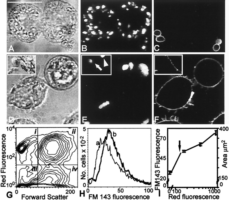
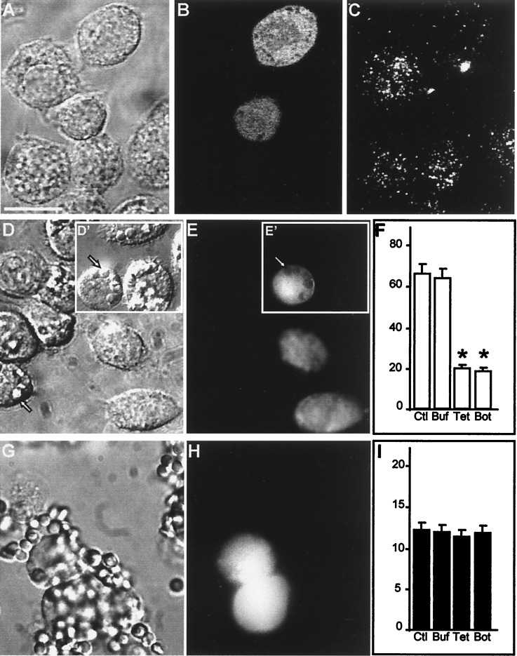
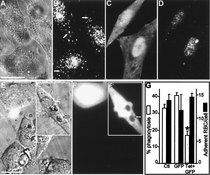
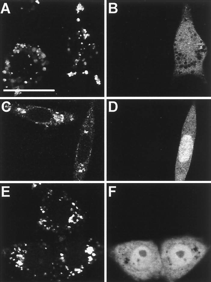
Phagosomes are generally believed to form by gradual apposition of the plasma membrane of leukocytes onto the surface of invading microorganisms. The internalization of the encapsulated particle is therefore predicted to reduce the surface area of the phagocyte. Contrary to this prediction, we observed that phagocytosis is associated with a net increase in cell surface area, suggesting the concomitant occurrence of exocytosis. Selective cleavage of components of the secretory machinery by microinjection or transfection of bacterial neurotoxins induced a pronounced inhibition of phagocytosis. These observations indicate that vesicle-soluble N-ethylmaleimide-sensitive factor attachment protein receptor-mediated exocytosis of endomembranes is essential for optimal completion of particle internalization during phagocytosis.


Figures

Figure 1
Figure 1
Changes in macrophage surface area after phagocytosis of OPZ. (A–C) J774 cells were allowed to interact with biotinylated, Texas red-labeled OPZ. Extracellular particles were identified by treatment with avidin-FITC. (A) Bright field (Nomarski) image. Bar, 10 μm. (B) Red fluorescence (Texas red emission) of cells in A as analyzed by confocal fluorescence microscopy, indicating the location of OPZ. (C) Green fluorescence (avidin-FITC emission) of cells in A, identifying extracellular OPZ. (D–F) J774 cells were allowed to internalize Texas red-labeled OPZ as in A, treated with the dye FM-143 at 4°C (which provides a measure of the surface membrane), and then examined by confocal microscopy. (D) Bright field image; inset shows an extracellular particle (arrow) and an intracellular particle (arrowhead). (E) Red (Texas red) fluorescence of cells in D indicating the location of OPZ. (F) Green (FM-143) fluorescence indicating surface labeling of cell only. (G) Analysis of association of OPZ with J774 cells by flow cytometry. The arbitrarily designated quadrants define the following populations: i, extracellular OPZ; ii, macrophages with associated OPZ; iii, debris; and iv, macrophages not associated with OPZ. (H) Frequency histogram of the fluorescence intensity of cell-associated FM-143, a measure of cell surface area. The lighter line (a) corresponds to cells in quadrant iv in G, which had no associated OPZ. The heavy line (b) corresponds to cells in quadrant ii in G, which had internalized OPZ. (I) Correlation between Texas red fluorescence (abscissa), which is proportional to the number of OPZ particles (fluorescence equivalent to one internalized particle shown by arrow) and the uptake of FM-143 (left ordinate), which is proportional to surface area (calculated from the volume of cells, assuming a smooth spherical surface; right ordinate). Data from five separate experiments.
Figure 2
Figure 2
Impaired phagocytosis after degradation of VAMP-2 in J774 cells. (A–C) J774 cells were injected with TeTx-LC in a solution containing fixable FITC-dextran as a marker of injection. After 5 h of incubation at 37°C, the cells were stained with antibody to VAMP-2. (A) Bright field micrograph. Bar, 10 μm. (B) Green fluorescence (Lucifer yellow emission) of the field shown in A identifying two injected cells. (C) VAMP-2 immunostaining in the cells shown in A. (D–F) J774 cells were injected with tetanus toxin and FITC-dextran as above, then assessed for their ability to perform phagocytosis of opsonized SRBCs. (D) Bright field micrograph. Inset (D′) shows cell injected with dextran alone. Arrow indicates location of representative internalized SRBCs. (E and E′) Green fluorescence emission of the cells shown in D. Arrowhead demonstrates the displacement of FITC-dextran by internalized the SRBCs. (F) Effect of toxin injection on phagocytosis. Data are means ± SE of 250 noninjected control (Ctl) cells, 175 buffer- (Buf) injected controls, 244 TeTx-LC- (Tet) injected cells, and 75 botulinum- (Bot) injected cells (asterisks indicate P < 0.05 vs. Ctl). (G–I) J774 cells were injected with TeTx-LC as above and then assessed for their ability to bind opsonized SRBCs. (G) Bright field micrograph. (H) Fluorescence emission of the cells shown in G identifying injected cells. (I) Effect of toxin injection on SRBC adherence to J774 cells. Data are means ± SE of 165 noninjected control (Ctl) cells, 64 buffer- (Buf) injected controls, 75 TeTx-LC- (Tet) injected cells, and 57 botulinum- (Bot) injected cells. Images are representative of five separate experiments.
Figure 3
Figure 3
Impaired phagocytosis after degradation of VAMP-2 in FcγRIIA-transfected CHO cells. (A and B) VAMP-2 immunostaining of CHO cells. (A) Nomarski image. (B) VAMP-2 immunostaining in the cells shown in A. Arrows indicate specific, vesicular staining. (C and D) FcγRIIA-expressing CHO cells were transiently transfected with cDNA-encoding TeTx-LC, along with cDNA for GFP as a transfection marker, and immunostained with antibody to VAMP-2. (C) Green (GFP) fluorescence identifying transfected cells. (D) VAMP-2 immunostaining of cells shown in C. (E and F) FcγRIIA-expressing CHO cells were cotransfected with TeTx-LC and GFP as above and allowed to internalize opsonized SRBCs. (E) Bright field image. Inset (E′) shows cells transfected with GFP alone. Arrowhead indicates location of representative internalized SRBCs. (F and F′) GFP fluorescence identifying transfected cells in the field shown in E. The arrowhead demonstrates the displacement of GFP by internalized SRBCs in cells transfected with GFP alone. (G) Effect of TeTx-LC expression on phagocytosis and adherence of SRBCs by FcγRIIA-CHO cells. Means ± SE of five individual experiments, with more than 200 cells per group per experiment. Asterisks indicate P < 0.05 vs. control (Ctl.). Bar, 10 μm.
Figure 4
Figure 4
Effect of tetanus toxin on endocytosis. (A and B) J774 cells were injected with TeTx-LC and fluorescein-dextran as an injection marker, incubated for 5 h at 37°C, and then allowed to internalize Texas red-dextran for 2 h at 37°C. (A) Typical confocal micrograph demonstrating Texas red fluorescence. (B) Fluorescein-dextran emission identifying injected cell. (C–F) FcγRIIA-expressing CHO cells were transiently transfected with cDNA for TeTx-LC and GFP and allowed to internalize either rhodamine-labeled transferrin (C and D) or tetramethylrhodamine B isothiocyanate-labeled aggregated human IgG (E and F) for 1 h at 37°C. (C and E) Confocal fluorescence micrograph illustrating the distribution of transferrin (C) or aggregated IgG (E). (D and F) GFP emission identifying transfected cells. Representative of three experiments, with at least 50 injected or transfected cells per experiment. Bar, 10 μm.

References

    1. Greenberg S, Silverstein S C. In: Phagocytosis. Paul W E, editor. New York: Raven; 1993. pp. 941–964.
    1. Desjardins M, Huber L A, Parton R G, Griffiths G. J Cell Biol. 1994;124:677–688. - PMC - PubMed
    1. Hackam D J, Rotstein O D, Zhang W J, Demaurex N, Woodside M, Tsai O, Grinstein S. J Biol Chem. 1997;272:29810–29820. - PubMed
    1. Griffin F M, Jr, Griffin J A, Leider J E, Silverstein S C. J Exp Med. 1975;142:1263–1282. - PMC - PubMed
    1. Griffin F M, Jr, Griffin J A, Silverstein S C. J Exp Med. 1976;144:788–809. - PMC - PubMed

Publication types

LinkOut - more resources