. 1998 Sep 29;95(20):11846-51.
doi: 10.1073/pnas.95.20.11846.

Parathyroid hormone-related protein is required for tooth eruption

Affiliations

Parathyroid hormone-related protein is required for tooth eruption

W M Philbrick et al. Proc Natl Acad Sci U S A. .

Abstract

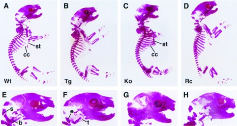
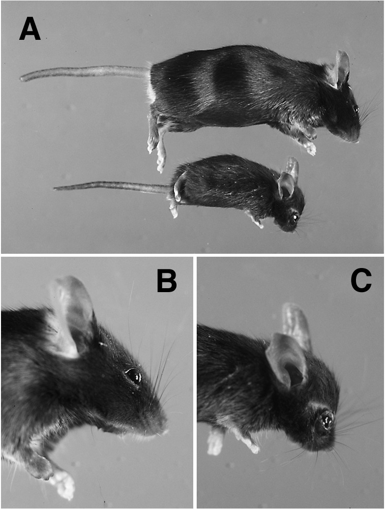
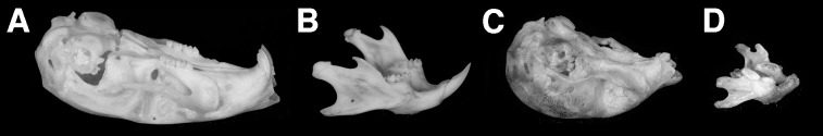
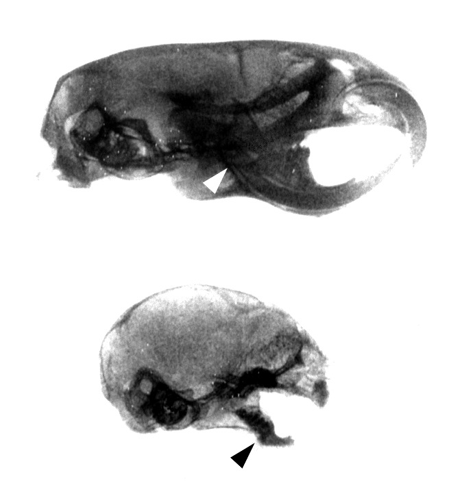
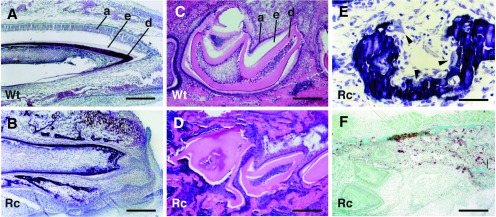
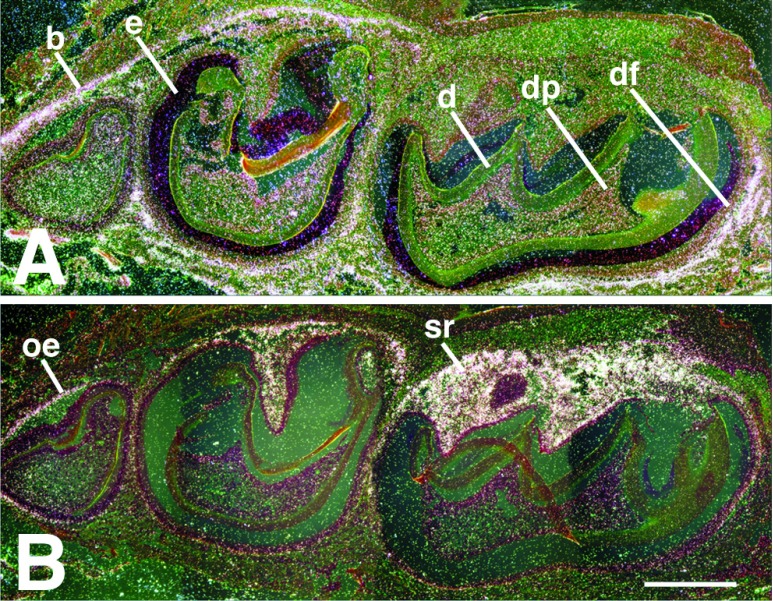
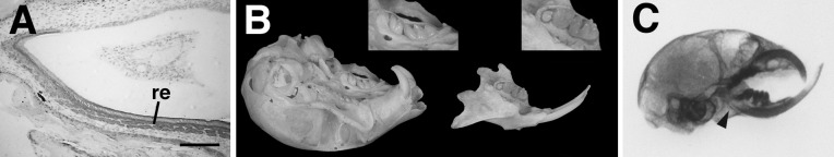
Parathyroid hormone (PTH)-related protein (PTHrP)-knockout mice die at birth with a chondrodystrophic phenotype characterized by premature chondrocyte differentiation and accelerated bone formation, whereas overexpression of PTHrP in the chondrocytes of transgenic mice produces a delay in chondrocyte maturation and endochondral ossification. Replacement of PTHrP expression in the chondrocytes of PTHrP-knockout mice using a procollagen II-driven transgene results in the correction of the lethal skeletal abnormalities and generates animals that are effectively PTHrP-null in all sites other than cartilage. These rescued PTHrP-knockout mice survive to at least 6 months of age but are small in stature and display a number of developmental defects, including cranial chondrodystrophy and a failure of tooth eruption. Teeth appear to develop normally but become trapped by the surrounding bone and undergo progressive impaction. Localization of PTHrP mRNA during normal tooth development by in situ hybridization reveals increasing levels of expression in the enamel epithelium before the formation of the eruption pathway. The type I PTH/PTHrP receptor is expressed in both the adjacent dental mesenchyme and in the alveolar bone. The replacement of PTHrP expression in the enamel epithelium with a keratin 14-driven transgene corrects the defect in bone resorption and restores the normal program of tooth eruption. PTHrP therefore represents an essential signal in the formation of the eruption pathway.


Figures

Figure 1
Figure 1
Staining of calcified tissue in neonatal mice with alizarin red S. From left to right are wild-type (A), col II-PTHrP transgenic (B), PTHrP-knockout (C), and rescued PTHrP-knockout (D) phenotypes. The PTHrP-knockout animals (C) display inappropriate ossification of the costal cartilage (cc) and along the length of the sternum (st). These abnormalities appear to be largely corrected in the rescued mice (D), in which ossification is confined to the sternebrae and does not extend to the costal cartilage. A magnified view of the rescued-knockout cranium (H) shows substantial correction of the accelerated mineralization of the endochondral bones of the cranial base seen in the knockout (G); s, supraoccipital; b, basioccipital; e, exoccipital; t, tympanic ring. The tips of the emerging mandibular incisors can be discerned in both the normal (E) and transgenic (F) animals, but not in the PTHrP-knockout (G) or rescued-knockout (H) mice. Wt, wild type; Tg, transgenic; Ko, knockout; Rc, rescued-knockout.
Figure 2
Figure 2
Gross appearance of rescued PTHrP-knockout mice. (A) Relative size of a rescued-knockout mouse at 8 weeks of age (below) as compared with a wild-type littermate (above). Foreshortening of the limbs is apparent by this age. The cranial chondrodystrophy in the rescued-knockout mouse (C), as compared with a normal animal (B), includes doming of the calvarium, frontal bossing, and a flattened snout.
Figure 3
Figure 3
Comparison of skulls from adult wild-type mice (A and B) and rescued PTHrP-knockouts (C and D). External incisors and molars are clearly absent from both the maxilla (compare A and C) and mandible (compare B and D) in these animals. The lack of functional dentition in the rescued-knockout is superimposed on the underlying features of the cranial chondrodystrophy, which includes foreshortening of the maxilla and mandible.
Figure 4
Figure 4
Radiographic analysis of tooth eruption in wild-type littermates (Upper) and rescued PTHrP-knockout mice (Lower) at 2 weeks of age. Normal eruption of incisors and molars (Upper) does not occur in the rescued-knockout mouse (Lower); the radiographic densities apparent in the mandible and maxilla of the rescued-knockouts represent impacted teeth. Also, the root of the lower incisor extends under the molars to M3 in the normal mandible (arrowhead, Upper), whereas the incisor root in the rescued-knockout fails to reach M1 (arrowhead, Lower), a finding typical of osteopetrosis.
Figure 5
Figure 5
Tooth histology. (A and B) Sagittal, nondecalcified sections of incisors from a rescued PTHrP-knockout neonate (B) and a wild-type littermate (A) stained with toluidine blue. A well developed ameloblast layer (a) is readily apparent on the labial aspect of the normal tooth (A), but is lacking in the mutant (B), which is crowded by the surrounding alveolar bone. The dark dentin layer (d) is intact but the adjacent enamel (e) is absent from the labial surface of the incisor (the space between the ameloblast layer and the enamel in A is a sectioning artifact). (C and D) Mandibles from 1-week-old wild-type (C) and rescued-knockout mice (D) were decalcified and then sectioned sagittally through the molar crypt. Distortion of the teeth in the rescued-knockout animal caused by progressive impaction is readily apparent. The space between the ameloblast (a) and dentin (d) layers defines the area occupied by the enamel (e) that has been removed through decalcification. (E) Magnification of an nondecalcified sagittal section through the region between the incisor and the first molar of a mandible from a rescued-knockout animal. Osteoclasts (arrowheads) can be seen attached to the surfaces of the alveolar bone. (F) Coronal sections of mandibles from neonatal rescued-knockout animals were stained for tartrate-resistant acid phosphatase activity. Tartrate-resistant acid phosphatase-positive cells (stained brown) are present in the alveolar bone immediately adjacent to the lateral aspect of the molars in a pattern indistinguishable from that found in wild-type littermates (not shown). Wt, wild type; Rc, rescued-knockout. [Bar = 64 μm in (A and B); 32 μm (C, D, and F), and 16 μm (E).]
Figure 6
Figure 6
In situ hybridization of a normal mandible sectioned sagittally through the molar crypt and hybridized with murine probes for the PTH/PTHrP receptor (A) and PTHrP (B). The receptor is found principally in bone (b), but can also be detected in the dental mesenchyme, both in the dental pulp (dp) and the dental follicle layer (df). PTHrP localizes to the outer enamel epithelium (oe) and the stellate reticulum (sr). Some enamel (e) remains after decalcification; d, dentin. [The scale bar represents 0.12 mm.]
Figure 7
Figure 7
Dental phenotype of doubly rescued PTHrP-knockout mice. (A) Immunohistochemical staining for K14 (dark gray) in a sagittal section from a normal neonatal incisor localizes to the reduced enamel epithelium (re) on the labial surface. (B) Eruption of both incisors and molars (enlarged in Insets) is readily apparent in the maxilla and mandible of an adult doubly rescued animal. (C) A sagittal radiograph of an adult doubly-rescued PTHrP-knockout mouse clearly reveals the malocclusion of the incisors in the rescued-knockout caused by the underlying chondrodystrophy. Note the restoration of root extension in the lower incisor (arrowhead). (Bar = 27 μm.)

References

    1. Broadus A E, Stewart A F. In: The Parathyroids. Bilezekian J P, Levine M A, Marcus R, editors. New York: Raven; 1994. pp. 259–294.
    1. Jüppner H, Abou-Samra A-B, Freeman M, Kong X F, Schipani E, Richards J, Kolakowski L F, Hock J, Potts J T, Kronenberg H M, et al. Science. 1991;254:1024–1026. - PubMed
    1. Abou-Samra A-B, Jüppner H, Force T, Freeman M W, Kong X-F, Schipani E, Urena P, Richards J, Bonventre J V, Potts J T, Jr, Kronenberg H M, et al. Proc Natl Acad Sci USA. 1992;89:2732–2736. - PMC - PubMed
    1. Campos R V, Asa S L, Drucker D J. Cancer Res. 1991;51:6351–6357. - PubMed
    1. Lee K, Deeds J D, Segre G V. Endocrinology. 1995;136:453–463. - PubMed

Publication types

MeSH terms

Substances

LinkOut - more resources