. 1998 Sep 29;95(20):11863-8.
doi: 10.1073/pnas.95.20.11863.

The PEBP2betaMYH11 fusion created by Inv(16)(p13;q22) in myeloid leukemia impairs neutrophil maturation and contributes to granulocytic dysplasia

Affiliations

The PEBP2betaMYH11 fusion created by Inv(16)(p13;q22) in myeloid leukemia impairs neutrophil maturation and contributes to granulocytic dysplasia

S C Kogan et al. Proc Natl Acad Sci U S A. .

Abstract

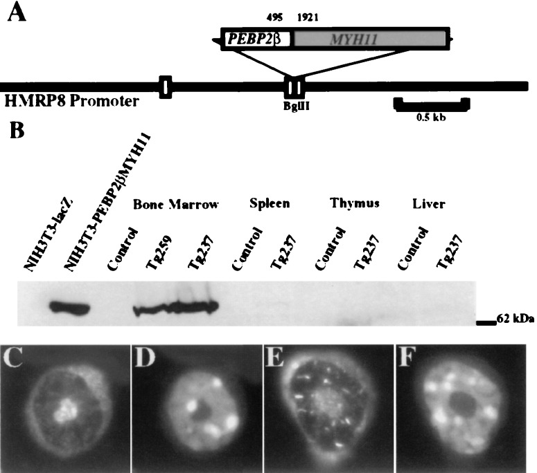
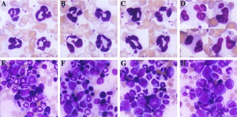
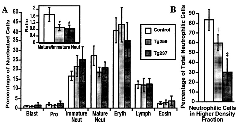
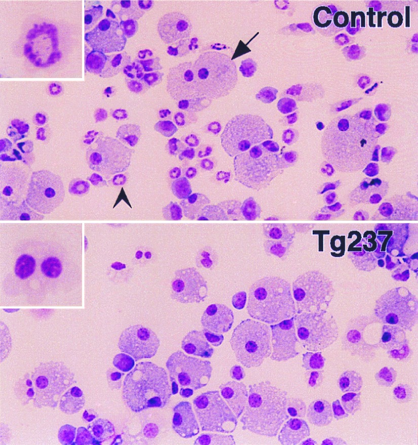
Chromosomal translocations involving the genes encoding the alpha and beta subunits of the Pebp2/Cbf transcription factor have been associated with human acute myeloid leukemia and the preleukemic condition, myelodysplasia. Inv(16)(p13;q22) fuses the gene encoding the beta subunit of Pebp2 to the MYH11 gene encoding a smooth muscle myosin heavy chain (Smmhc). To examine the effect of the inv(16)(p13;q22) on myelopoiesis, we used the hMRP8 promoter element to generate transgenic mice expressing the Pebp2betaSmmhc chimeric fusion protein in myeloid cells. Neutrophil maturation was impaired in PEBP2betaMYH11 transgenic mice. Although the transgenic mice had normal numbers of circulating neutrophils, their bone marrow contained increased numbers of immature neutrophilic cells, which exhibited abnormal characteristics. In addition, PEBP2betaMYH11 inhibited neutrophilic differentiation in colonies derived from hematopoietic progenitors. Coexpression of both PEBP2betaMYH11 and activated NRAS induced a more severe phenotype characterized by abnormal nuclear morphology indicative of granulocytic dysplasia. These results show that PEBP2betaMYH11 can impair neutrophil development and provide evidence that alterations of Pebp2 can contribute to the genesis of myelodysplasia.


Figures

Figure 1
Figure 1
(A) Map of the transgene. The PEBP2βMYH11 cDNA inserted into the BglII site of the human MRP8 promoter cassette corresponded to the most common inv(16) breakpoint. The nucleotide locations of the breakpoint within the coding sequences of PEBP2β and MYH11 are shown. White boxes represent noncoding portions of MRP8 exons. Solid lines represent 5′ and 3′ flanking sequences and intron 1 of MRP8. (B) Expression of the transgene. Whole-cell lysates were subjected to denaturing PAGE and Western blotting using a rabbit polyclonal anti-Pebp2β antiserum. The signal at 70 kDa corresponds to the Pebp2βSmmhc protein. The first two lanes are lysates of NIH 3T3 cells that contained retroviral vectors expressing either a lacZ or a PEBP2βMYH11 cDNA. The remaining lanes are lysates of tissues from control FVB/N mice or transgenic mice of lines Tg259 or Tg237. (CF) Immunofluorescence of bone marrow neutrophilic cells. (C and D) Control. (E and F) Tg237. (C and E) Anti-Pebp2β antiserum. (D and F) Hoechst 33258. (Original magnification, ×250.)
Figure 2
Figure 2
Aberrant neutrophilic maturation in PEBP2βMYH11 transgenic mice. (A and E) Samples from control mice. (B and F) Samples from PEBP2βMYH11 transgenic mice (line Tg237). (C and G) Samples from NRASG12D transgenic mice (line Tg7). (D and H) Samples from PEBP2βMYH11/NRASG12D double transgenic mice. (AD) Peripheral blood neutrophils. (EH) Bone marrow. (Original magnification, ×250.)
Figure 3
Figure 3
(A) Bone marrow differential cell counts. Differential cell counts (total of 400 cells each) were performed on bone marrow smears. Data are shown as arithmetic means ± SD. Pro, promyelocytes; Immature Neut, neutrophilic myelocytes and metamyelocytes; Mature Neut, neutrophilic band, mature ring, and polymorphonuclear forms; Eryth, nucleated erythroid cells; Lymph, lymphocytes; Eosin, eosinophilic cells. Control, n = 6; Tg259, n = 6; Tg237, n = 6. (Inset) Ratios of mature to immature neutrophilic cells. ∗ indicates a significant shift toward immature cells is present in the bone marrow of the transgenic mice, P = 0.004. (B) Density of bone marrow neutrophilic cells. Bone marrow cells were separated into low and high density fractions on a Percoll step gradient. Total numbers of cells present in each fraction were determined. Differential cell counts (total of 100 cells each) were performed on cytospins of each fraction, with cells morphologically identified as neutrophilic (neutrophilic myelocytes, metamyelocytes, ring, band, and polymorphonuclear forms) or nonneutrophilic. Total cell counts and differential counts were used to calculate the mean percentages of neutrophilic cells present in the high density fractions. Data are shown as arithmetic means ± SD. Control, n = 7; Tg259, n = 5; Tg237, n = 6. †, Control vs. Tg259, P = 0.0006. ‡, Control vs. Tg237, P = 0.00002; Tg259 vs. Tg237, P = 0.002.
Figure 4
Figure 4
Morphology of bone marrow cells cultured in vitro. Bone marrow cells were grown in semisolid methylcellulose medium in the presence of G-CSF at 2.5 × 10⁵ cells/ml for 7 days, and cytospins of harvested cells were prepared. Numerous macrophages (arrow) and neutrophilic cells (arrowhead) were observed in the control samples. Neutrophilic cells were decreased in the samples from transgenic mice. (Insets) Neutrophil morphology. Neutrophils derived in vitro from the bone marrows of transgenic mice exhibited atypical morphology. (Original magnification, ×250.)

References

    1. Pedersen-Bjergaard J, Rowley J D. Blood. 1994;83:2780–2786. - PubMed
    1. Liu P, Tarle S A, Hajra A, Claxton D F, Marlton P, Freedman M, Siciliano M J, Collins F S. Science. 1993;261:1041–1044. - PubMed
    1. Le Beau M M, Larson R A, Bitter M A, Vardiman J W, Golomb H M, Rowley J D. N Engl J Med. 1983;309:630–636. - PubMed
    1. Langabeer S E, Walker H, Gale R E, Wheatley K, Burnett A K, Goldstone A H, Linch D C. Br J Haematol. 1997;96:736–739. - PubMed
    1. Speck N A, Stacy T. In: Critical Reviews in Eukaryotic Gene Expression. Stein G S, Stein J L, Lian J B, editors. New York: Begell House; 1995. pp. 337–364.

Publication types

MeSH terms