. 1998 Sep 29;95(20):11869-74.
doi: 10.1073/pnas.95.20.11869.

Altered surfactant homeostasis and alveolar type II cell morphology in mice lacking surfactant protein D

Affiliations

Altered surfactant homeostasis and alveolar type II cell morphology in mice lacking surfactant protein D

C Botas et al. Proc Natl Acad Sci U S A. .

Abstract

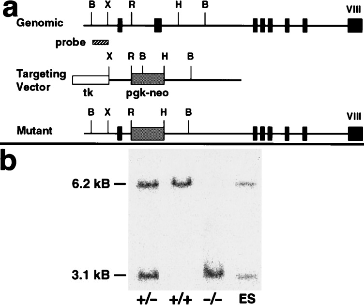
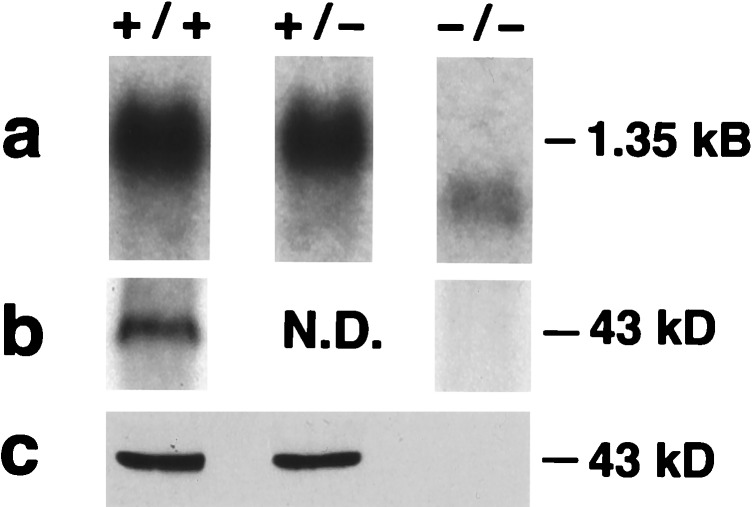
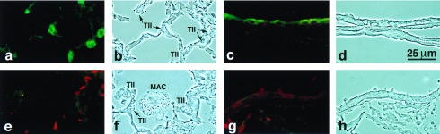
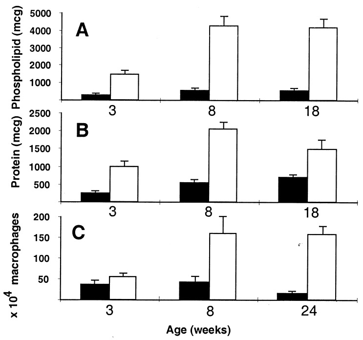
Surfactant protein D (SP-D) is one of two collectins found in the pulmonary alveolus. On the basis of homology with other collectins, potential functions for SP-D include roles in innate immunity and surfactant metabolism. The SP-D gene was disrupted in embryonic stem cells by homologous recombination to generate mice deficient in SP-D. Mice heterozygous for the mutant SP-D allele had SP-D concentrations that were approximately 50% wild type but no other obvious phenotypic abnormality. Mice totally deficient in SP-D were healthy to 7 months but had a progressive accumulation of surfactant lipids, SP-A, and SP-B in the alveolar space. By 8 weeks the alveolar phospholipid pool was 8-fold higher than wild-type littermates. There was also a 10-fold accumulation of alveolar macrophages in the null mice, and many macrophages were both multinucleated and foamy in appearance. Type II cells in the null mice were hyperplastic and contained giant lamellar bodies. These alterations in surfactant homeostasis were not associated with detectable changes in surfactant surface activity, postnatal respiratory function, or survival. The findings in the SP-D-deficient mice suggest a role for SP-D in surfactant homeostasis.


Figures

Figure 1
Figure 1
Targeting strategy for SP-D gene disruption and confirmation of the expected null mutation by genotype analysis. (a) The thin line depicts the mouse SP-D gene, with the eight exons indicated by boxes. Pgk-neo replaced all of exon 2 that contains the translation start site for SP-D. The 600-bp probe used for Southern blot screening is located 5′ to the targeting vector. B, BstXI; r, EcoRI; H, HindIII; X, XbaI. (b) Southern blot of tail genomic DNA from wild type (+/+), heterozygous (+/−), and SP-D null (−/−) mice and embryonic stem clone DNA after BstXI digestion. The 6.2-kb BstXI band is the normal allele and the 3.1-kb BstXI band the mutant allele.
Figure 2
Figure 2
Analysis of SP-D mRNA (a) and protein in the lungs (b) and BAL (c) of null mice. (a) Northern blot of lung RNA demonstrating the absence of a 1.35-kb full-length mRNA for SP-D in the lungs of −/− mice. The low level 1-kb mRNA lacking exon 2 is visible. (b) Western blot of lung tissue demonstrating the absence of SP-D in the −/− lung. (c) Western blot of BAL demonstrating the absence of SP-D in −/− BAL.
Figure 3
Figure 3
SP-D immunohistochemistry in null and wild-type mice. SP-D is detected in type II cells (a) and Clara cells (c) of wild-type mice but is not detected in SP-D null mice (e–h). Foamy alveolar macrophages are visible in the alveolus of the null (f).
Figure 4
Figure 4
BAL analysis of SP-D +/+ (solid bars) and SP-D −/− (open bars). (A) Total BAL phospholipid per mouse at 3, 8, and 18 weeks (n = 8 for each genotype at each age; P < 0.001). (B) Total BAL protein per mouse at 3, 8, and 18 weeks (n = 8 for each genotype at each age; P < 0.01). (C) Number of alveolar macrophages per mouse at 3, 8, and 24 weeks (n = 4 to 7 for each genotype at each age; P < 0.05 at 3 and 24 weeks). Error bars indicate the SEM.
Figure 5
Figure 5
Effect of bubble oscillation on quasistatic surface tension–area isotherms of surfactants from null and wild-type mice. Open symbols (null) and solid symbols (wild type) represent averages of five preparations. Error bars indicate the standard errors of averages.
Figure 6
Figure 6
Lung histology demonstrating progressive accumulation of surfactant lipids in the alveolar space, foamy macrophages, and peribronchial infiltrate in SP-D null mice. (ac) Sections from wild-type mice at 3, 8, and 18 weeks, respectively. (df) Sections from SP-D null mice at 3, 8, and 18 weeks, respectively.
Figure 7
Figure 7
Electron microscopy of an alveolar region demonstrating massive accumulation of intraalveolar surfactant and giant lamellar bodies (c) and stuffed macrophages (b) in the SP-D null mouse compared with the wild-type littermate (a). All micrographs are the same magnification. (Bar = 10 μm in c.)
Figure 8
Figure 8
Cytospins of BAL cell pellets at 3 weeks (a and b) and 24 weeks (c and d) in wild-type (a and c) and SP-D null mice (b and d). The increased size of the macrophages in the null mice is visible. The arrows indicate multinucleated macrophages from null mice.

References

    1. Persson A, Chang D, Rust K, Moxley M, Longmore W, Crouch E. Biochemistry. 1989;28:6361–6367. - PubMed
    1. Wong C J, Akiyama J, Allen L, Hawgood S. Pediatr Res. 1996;39:930–937. - PubMed
    1. Fisher J H, Mason R. Am J Respir Cell Mol Biol. 1995;12:13–18. - PubMed
    1. Hoppe H J, Reid K B. Protein Sci. 1994;3:1143–1158. - PMC - PubMed
    1. Akiyama J, Poulain F, Vanderbilt J, Hawgood S. Am J Respir Crit Care Med. 1998;157:A561. (abstr.).

Publication types

MeSH terms