

Opposing actions of prostaglandins and oxytocin determine the onset of murine labor
- PMID: 9751758
- PMCID: PMC21733
- DOI: 10.1073/pnas.95.20.11875
Opposing actions of prostaglandins and oxytocin determine the onset of murine labor
Abstract
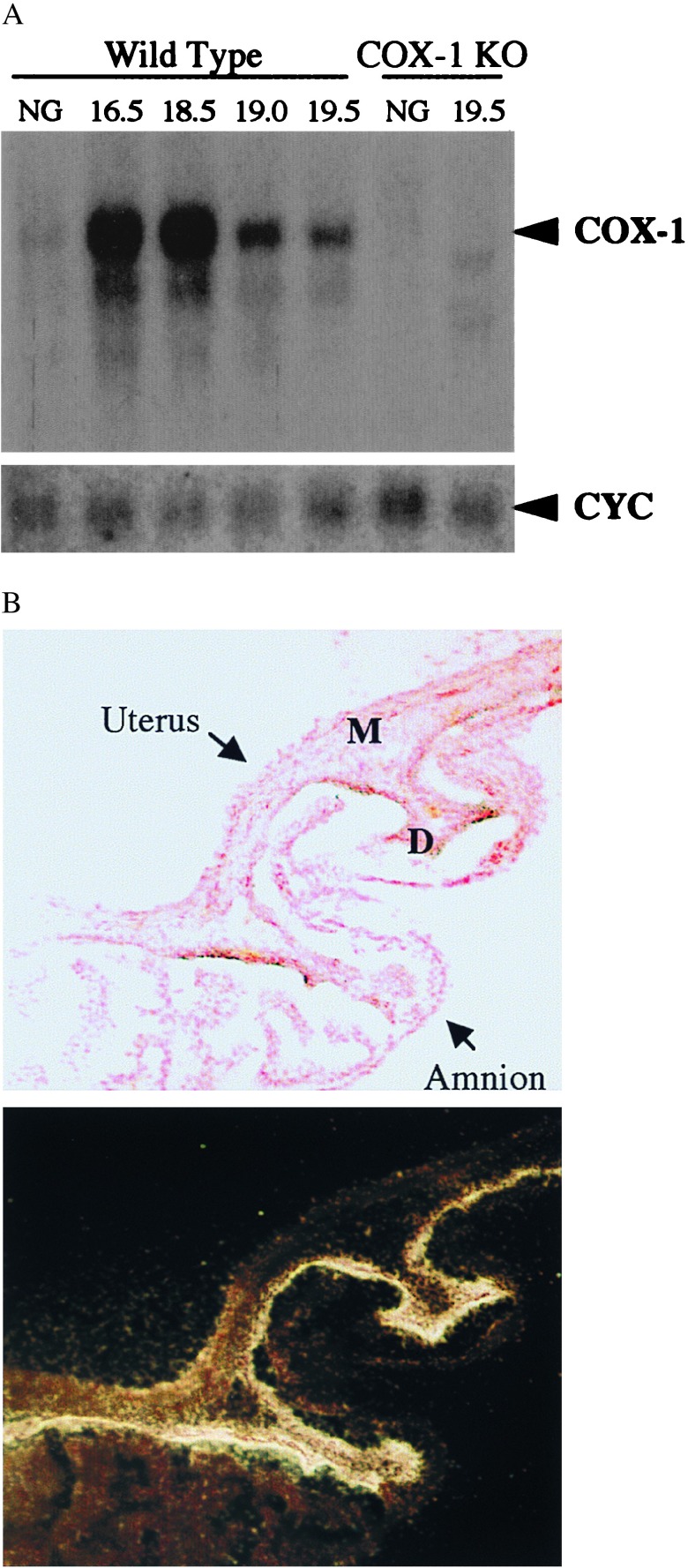
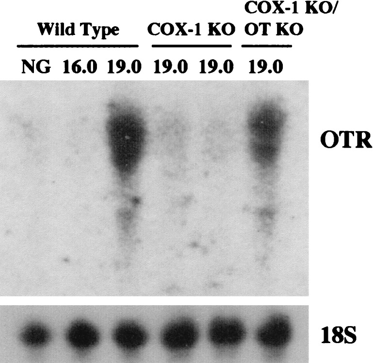
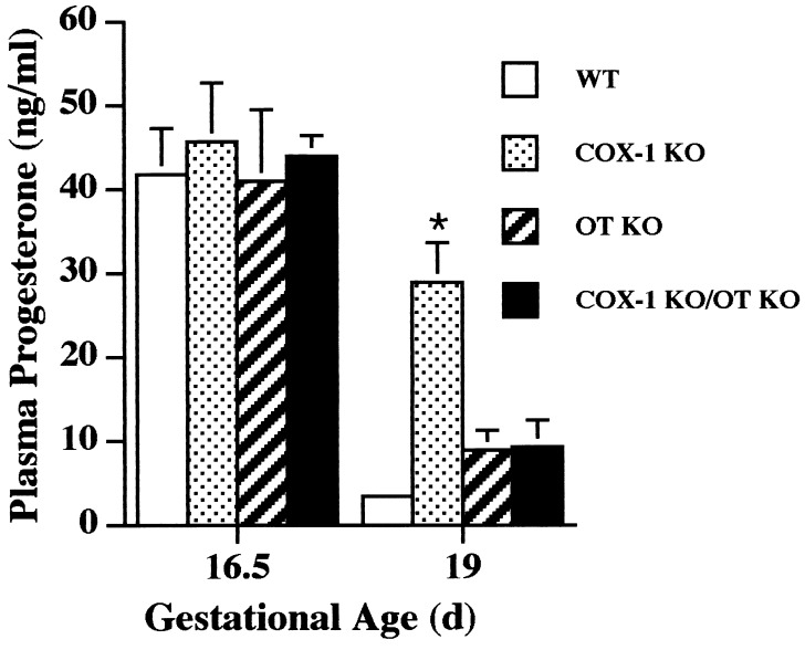
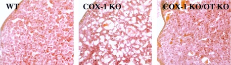
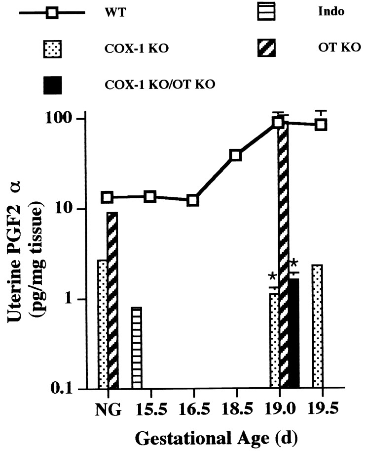
Prostaglandins (PGs) have been recently proven essential for parturition in mice. To dissect the contributions of the two cyclooxygenase (COX) isoforms to the synthesis of PGs during pregnancy, we have characterized the parturition phenotype of COX-1-deficient mice. We find that mice with targeted disruption of the COX-1 gene have delayed parturition resulting in neonatal death. Results of matings of COX-1-deficient females with COX-1 intact males, and blastocyst transfer of COX-1-deficient or -intact embryos into wild-type foster mothers, proved necessity and sufficiency of maternal COX-1 for the normal onset of labor. COX-1 expression is induced in gravid murine uterus and by in situ hybridization; this induction is localized to the decidua. Measurement of uterine PGs further confirmed that COX-1 accounted for the majority of PGF2alpha production. To evaluate the interaction of PGs with oxytocin during murine labor, we generated mice deficient in both oxytocin and COX-1. Surprisingly, the combined oxytocin and COX-1-deficient mice initiated labor at the normal time. COX-1-deficient mice demonstrated impaired luteolysis, as evidenced by elevated serum progesterone concentration and ovarian histology late in gestation, and delayed induction of uterine oxytocin receptors. In contrast, simultaneous oxytocin and COX-1 deficiency restored the normal onset of labor by allowing luteolysis in the absence of elevated PGF2alpha production. These findings demonstrate that COX-1 is essential for normal labor in the mouse, with a critical function being to overcome the luteotrophic action of oxytocin in late gestation.
Figures
References
-
- Crowley P, Chalmers M J N C. Br J Obstet Gynaecol. 1990;97:11–25. - PubMed
-
- NIH Concensus Development Panel on the Effect of Corticosteroids for Fetal Maturation on Perinatal Outcomes. J Am Med Assoc. 1995;273:413–418. - PubMed
-
- Uozumi N, Kume K, Nagase T, Nakatani N, Ishii S, Tashiro F, Komagata Y, Maki K, Ikuta K, Ouchi Y, et al. Nature (London) 1997;390:618–622. - PubMed
-
- Sugimoto Y, Yamasaki A, Segi E, Tsuboi K, Aze Y, Nishimura T, Oida H, Yoshida N, Tanaka T, Katsuyama M, et al. Science. 1997;277:681–683. - PubMed
-
- Larcher A, Neculcea J, Breton C, Arslan A, Rozen F, Russo C, Zingg H H. Endocrinology. 1995;136:5350–5356. - PubMed
Publication types
MeSH terms
Substances
Grants and funding
LinkOut - more resources
Full Text Sources
Molecular Biology Databases
