

Effect of vitamin A supplementation on rhodopsin mutants threonine-17 --> methionine and proline-347 --> serine in transgenic mice and in cell cultures
- PMID: 9751768
- PMCID: PMC21743
- DOI: 10.1073/pnas.95.20.11933
Effect of vitamin A supplementation on rhodopsin mutants threonine-17 --> methionine and proline-347 --> serine in transgenic mice and in cell cultures
Abstract
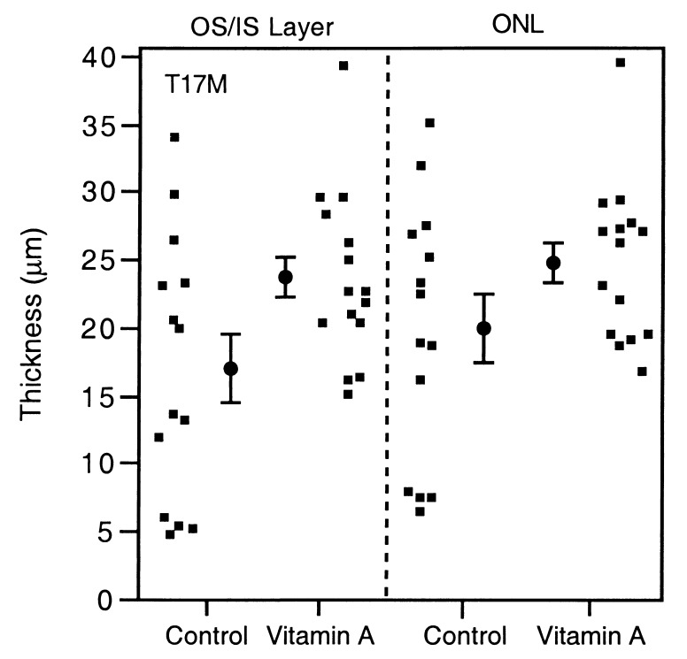
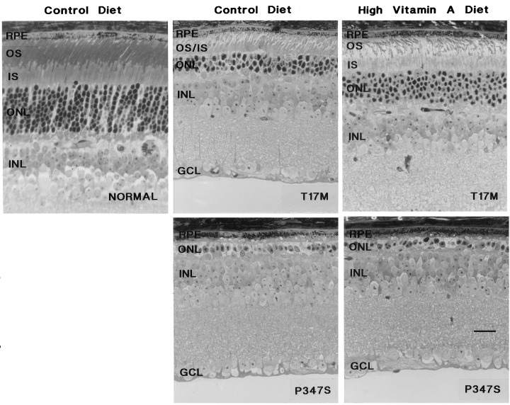
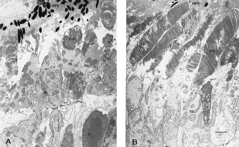
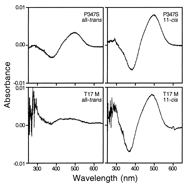
A therapeutic effect of vitamin A supplementation on the course of photoreceptor degeneration, previously reported for patients with retinitis pigmentosa, was tested in two transgenic mouse models of this disease, each carrying a dominant rhodopsin mutation. The threonine-17 --> methionine (T17M) mutation is a class II rhodopsin mutation, characterized by a thermal instability/folding defect and minimal regeneration with the chromophore. The proline-347 --> serine (P347S) mutation belongs to class I, comprised of a smaller number of mutations that exhibit no recognized biochemical abnormality in vitro. In the present study, each of the two mouse models was fed a diet containing 2.5 mg of vitamin A palmitate (control) or 102.5 mg of vitamin A palmitate (high vitamin A) per kilogram of diet. Dark-adapted, full-field electroretinograms showed that the high vitamin A diet significantly reduced the rate of decline of a-wave and b-wave amplitudes in the T17M mice but had no significant effect on the decline of electroretinogram amplitude in the P347S mice. Correspondingly, histologic evaluation revealed that the treatment was associated with significantly longer photoreceptor inner and outer segments and a thicker outer nuclear layer in the T17M mice but had no effect on photoreceptor morphology in the P347S mice. In a separate series of experiments, the instability defect of the T17M mutant opsin expressed in vitro was partially alleviated by inclusion of 11-cis-retinal in the culture media. These results show that vitamin A supplementation slows the rate of photoreceptor degeneration caused by a class II rhodopsin mutation. Vitamin A supplementation may confer therapeutic benefit by stabilizing mutant opsins through increased availability of the chromophore.
Figures


References
-
- Berson E L. Invest Ophthalmol Visual Sci. 1993;34:1659–1676. - PubMed
-
- Berson E L, Rosner B, Sandberg M A, Hayes K C, Nicholson B W, Weigel-DiFranco C, Willett W. Arch Ophthalmol. 1993;111:761–772. - PubMed
-
- Daiger S, Sullivan L, Rodriguez J. Behav Brain Sci. 1995;18:452–467.
-
- Sung C H, Davenport C M, Nathans J. J Biol Chem. 1993;268:26645–26649. - PubMed
Publication types
MeSH terms
Substances
Grants and funding
LinkOut - more resources
Full Text Sources
Other Literature Sources
Medical
Molecular Biology Databases
