. 1998 Sep 29;95(20):11975-80.
doi: 10.1073/pnas.95.20.11975.

Inhibition of the p44/42 MAP kinase pathway protects hippocampal neurons in a cell-culture model of seizure activity

Affiliations

Inhibition of the p44/42 MAP kinase pathway protects hippocampal neurons in a cell-culture model of seizure activity

B Murray et al. Proc Natl Acad Sci U S A. .

Abstract

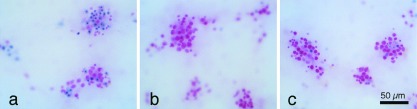
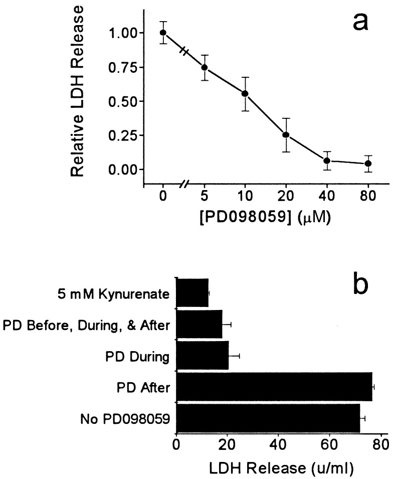
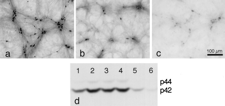
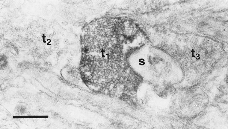
Excessive release of glutamate and the subsequent influx of calcium are associated with a number of neurological insults that result in neuronal death. The calcium-activated intracellular signaling pathways responsible for this excitotoxic injury are largely unknown. Here, we report that PD098059, a selective inhibitor of the calcium-activated p44/42 mitogen-activated protein kinase (MAP kinase) pathway, reduces neuronal death in a cell-culture model of seizure activity. Dissociated hippocampal neurons grown chronically in the presence of kynurenate, a broad spectrum glutamate-receptor antagonist, and elevated amounts of magnesium exhibit intense seizure-like activity after the removal of these blockers of excitatory synaptic transmission. A 30-min removal of the blockers produced extensive neuronal death within 24 h as assayed by the uptake of trypan blue and the release of lactate dehydrogenase. Phospho-p44/42 MAP kinase immunoreactivity after 30 min of seizure-like activity was present in many neuronal somata and dendrites as well as some synaptic terminals, consistent with both the presynaptic and postsynaptic effects of this pathway. The addition of PD098059 (40 microM; EC50 = 10 microM) during a 30-min washout of synaptic blockers inhibited the phosphorylation of p44/42 MAP kinase and reduced both the trypan-blue staining (n = 13) and the release of lactate dehydrogenase (n = 16) by 73% +/- 18% and 75% +/- 19% (mean +/- SD), respectively. The observed neuroprotection could be caused by an effect of PD098059 on seizure-like events or on downstream signaling pathways activated by the seizure-like events. Either possibility suggests a heretofore unknown function for the p44/42 MAP kinase pathway in neurons.


Figures

Figure 1
Figure 1
Trypan-blue staining, a marker of neuronal injury, 24 h after a 30-min washout of synaptic blockers. (a) Approximately half of the neurons stained for trypan blue after a 30-min washout of synaptic blockers. (b) Few neurons were stained for trypan blue in cultures that remained in a synaptic blocker (5 mM kynurenate) or (c) that were treated with 40 μM PD098059 during the washout of blockers. Neurons were counterstained with basic fuchsin (pink).
Figure 2
Figure 2
Release of LDH, a marker of neuronal injury, 24 h after a 30-min washout of synaptic blockers. (a) PD098059, at the concentrations indicated, was added 1 h before, during, and 24 h after a 30-min washout of kynurenate. PD098059 reduced the release of LDH with an EC50 of ≈10 μM, close to the IC50 for MEK1 inhibition (mean ± SEM, three to seven cultures per dose). (b) Adding PD098059 (40 μM) only during the washout of synaptic blockers (PD During) prevented the release of LDH and was as effective as adding it before, during and after the washout (PD Before, During, & After). PD098059 did not block the release of LDH if it was added only after the washout of synaptic blockers (PD After). In this experiment, PD098059 was only slightly less effective than 5 mM kynurenate in preventing the release of LDH (mean ± SEM, three cultures per condition).
Figure 3
Figure 3
Phospho-p44/42 MAP kinase immunohistochemistry after 30 min in EBSS without synaptic blockers or after 30 min in EBSS + 5 mM kynurenate. (a and c) After the washout of blockers, staining was prominent in the processes, many somata, and some nuclei. (b and d) In cultures that remained in synaptic blockers, faint staining of the processes and rare staining of the cell bodies were observed. Punctate staining that appeared to be intracellular was observed in some neurons that remained in synaptic blockers (b). Smaller punctate staining that appeared to be on the surface of the neurons was more prominent after the washout of blockers and likely indicates phosphorylation of p44/42 MAP kinase in synaptic endings. Neurons were counterstained with basic fuchsin (pink).
Figure 4
Figure 4
Effect of PD098059 on the phosphorylation of p44/42 MAP kinase after the removal of synaptic blockers. (a) In this plating, ≈60% of neurons were phospho-p44/42 MAP kinase-immunoreactive after 30 min in EBSS without synaptic blockers. (b) Sister cultures transferred to EBSS + 5 mM kynurenate showed less staining (≈13% of neurons). (c) Staining in cultures transferred to EBSS + 40 μM PD098059 was almost undetectable. Neurons were not counterstained. (d) Western blot showing the course of p44/42 MAP kinase phosphorylation over time after the removal of synaptic blockers and the inhibition by kynurenate and PD098059. The lengths of time that synaptic blockers were removed are: lane 1, 0 min; lane 2, 15 min; lane 3, 30 min; lane 4, 60 min; lane 5, 30 min (with 5 mM kynurenate); and lane 6, 30 min (with 40 μM PD098059). The phosphorylation at 0 min appears to be caused by the triplicate wash in EBSS + kynurenate immediately before cell lysis and assay (see text).
Figure 5
Figure 5
Electron micrograph of phospho-p44/42 MAP kinase immunoreactivity after 30 min in EBSS without synaptic blockers. Electron-dense label can be seen in a vesicle-filled terminal (t1) in close apposition to a small, unlabeled spine (s). The flattened disk-shaped structures in the spine are characteristic of the spine apparatus. Two other unlabeled terminals are visible (t2, t3). This section was not stained with uranyl acetate or lead citrate. (Bar = 0.5 μm.)

References

    1. Girault J-A. Neurochem Int. 1993;23:1–25. - PubMed
    1. Davis R J. J Biol Chem. 1993;268:14553–14556. - PubMed
    1. Seger R, Krebs E G. FASEB J. 1995;9:726–735. - PubMed
    1. Boulton T G, Nye S H, Robbins D J, Ip N Y, Radziejewska E, Morgenbesser S D, DePinho R A, Panayotatos N, Cobb M H, Yancopoulos G D. Cell. 1991;65:663–675. - PubMed
    1. Segal R A, Greenberg M E. Annu Rev Neurosci. 1996;19:463–489. - PubMed

Publication types

MeSH terms

Substances