Review
. 1998 Sep 29;95(20):11993-8.
doi: 10.1073/pnas.95.20.11993.

Interpreting functional imaging studies in terms of neurotransmitter cycling

Affiliations
Review

Interpreting functional imaging studies in terms of neurotransmitter cycling

R G Shulman et al. Proc Natl Acad Sci U S A. .

Abstract

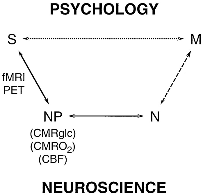
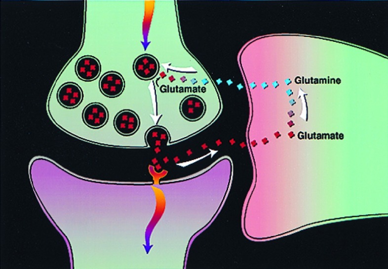
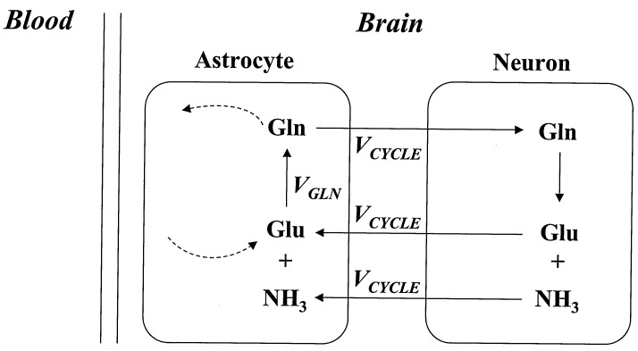
Functional imaging experiments, in particular positron-emission tomography and functional magnetic resonance imaging, can be analyzed either in psychological terms or on the basis of neuroscience. In the usual psychological interpretation, stimulations are designed to activate specific mental processes identified by cognitive psychology, which are then localized by the signals in functional imaging experiments. An alternate approach would be to analyze experiments in terms of the neurobiological processes responsible for the signals. Recent in vivo 13C NMR measurements of the glutamate-to-glutamine neurotransmitter cycling in rat and human brains facilitate a neuroscientific interpretation of functional imaging data in terms of neurobiological processes since incremental neurotransmitter flux showed a 1:1 stoichiometry with the incremental rate of glucose oxidation. Because functional imaging signals depend on brain energy consumption, a quantitative relationship can be established between the signal (S) and the specific neurochemical cerebral neurotransmitter activity (N) of glutamate-to-glutamine neurotransmitter cycling. The quantitation of neuronal activity proposed has implications for the psychological design and interpretation of functional imaging experiments. Measurements of the neurotransmitter cycling flux at rest in functional imaging experiments suggest that performing cognitive tasks and sensory stimulations increases neurotransmitter cycling by only 10-20%. Therefore it cannot be assumed that reference state activities are negligible, nor that they are constant during stimulation.


Figures

Figure 1
Figure 1
Schematic relations between the signal (S) obtained in functional imaging experiments and mental processes (M). In the usual experimental plan and interpretation, based on psychology, a direct relationship between S and M is assumed, as represented by the upper pathway. The definition of M is based on psychology, while the imaging experiment serves to localize and quantitate the brain activity identified with the process. The lower pathway, Neuroscience, assumes that M has a molecular and cellular basis, which is broken into three steps leading to S. The signal, S, in fMRI or PET experiments, is primarily a measure of the neurophysiological parameters (NP) of cerebral metabolic rate of glucose consumption (CMRglc), cerebral metabolic rate of oxygen consumption (CMRO2) or CBF. PET methods have been developed for measuring each of these three parameters separately, while fMRI signals respond to differences in the changes of CBF and CMRO2, whose quantitative relationships are being investigated. CMRO2 and CMRglc measure cerebral energy consumption, while ΔCMRO2 and ΔCMRglc measure its increment. The relation between (NP) the neurophysiological measure of energy consumption and neuronal activity (N) has been clarified by the 13C MRS experiments (–10). These recent findings allow measurements of S to be converted into measures of N, which places us squarely facing the unsolved “hard” problem of neuroscience, i.e., what is the relationship between M and N?
Figure 2
Figure 2
Proposed pathway of glutamate/glutamine neurotransmitter cycling between neurons and glia (22, 23), whose flux has been quantitated recently by 13C MRS experiments (9). Action potentials reaching the presynaptic neuron cause release of vesicular glutamate into the synaptic cleft, where it is recognized by glutamate receptors post-synaptically and is cleared by Na+ -coupled transport into glia. There it is converted enzymatically to glutamine, which passively diffuses back to the neuron and, after reconversion to glutamate, is repackaged into vesicles. The rate of the glutamate-to-glutamine step in this cycle (Vcycle), has been derived from recent 13C experiments (–10).
Figure 3
Figure 3
The experimental measure of VGln, the flux from glutamate to glutamine, includes, in addition to Vcycle, contributions from other pathways, as indicated by dashed lines, which have been shown to require small (≈10%) corrections to VGln to obtain Vcyle.
Figure 4
Figure 4
Experimental results of Vcycle and the rates of glucose oxidation measured simultaneously in each rat going from N2O/morphine to α-chloralose to pentobarbital (least active), at graded anesthesia. The best fit line gives CMRglc(ox) = 1.04 Vcycle + 0.10. The slope shows that each mole of neurotransmitter glutamate cycling requires the oxidation of one mole of glucose. The awake, resting state has CMRglc(ox) ≈0.8 μmol/min/g, which means that under this condition ≈85% of the brain energy consumption is dedicated to Vcycle (9).
Figure 5
Figure 5
Schematic representation of the signals obtained in functional imaging differencing experiments. PET measurements of CMRglc and CMRO2 directly measure S and (S + ΔS) under both conditions, although usually only the difference ΔS is used to localize and quantitate the task activation. On the other hand, fMRI signals include strong contributions in both conditions from pure imaging signals, so that in these experiments only ΔS is readily related to task activations.

References

    1. Raichle M E. In: Higher Functions of the Brain, Handbook of Physiology: The Nervous System. Plum F, Montcastle V, editors. Vol. 5. Washington, DC: Am. Physiol. Soc.; 1987. pp. 643–674.
    1. Sokoloff L, Reivich M, Kennedy C, Des Rosiers M H, Patlas C S, Pettigrew K D, Sakurada O, Shinohara M. J Neurochem. 1977;28:897–916. - PubMed
    1. Shulman R G, Blamire A M, Rothman D L, McCarthy G. Proc Natl Acad Sci USA. 1993;90:3127–3133. - PMC - PubMed
    1. Bandettini P A, Wong E C, Hinks R S, Tikofsky R S, Hyde J S. Magn Reson Med. 1992;25:390–397. - PubMed
    1. Raichle M E. Proc Natl Acad Sci USA. 1998;95:765–772. - PMC - PubMed

Publication types

LinkOut - more resources