

Altered thalamic response to levodopa in Parkinson's patients with dopa-induced dyskinesias
- PMID: 9751782
- PMCID: PMC21757
- DOI: 10.1073/pnas.95.20.12016
Altered thalamic response to levodopa in Parkinson's patients with dopa-induced dyskinesias
Abstract
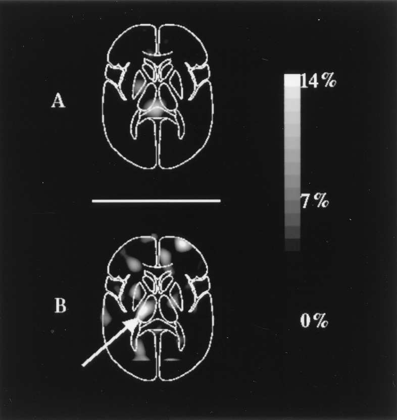
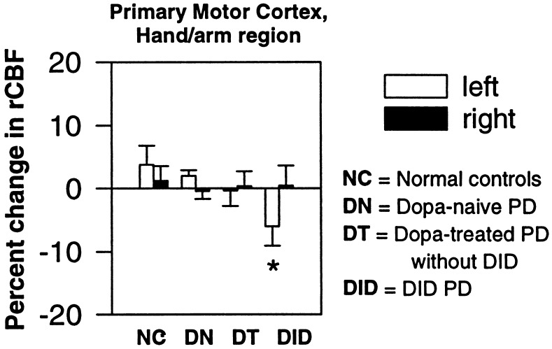
Parkinson's disease (PD) is a progressive neurologic condition characterized by tremor, slowness, stiffness, and unstable posture. Degeneration of dopamine-producing neurons in the substantia nigra causes PD. Treatment with levodopa, a precursor of dopamine, initially ameliorates the clinical manifestations of PD. However, chronic levodopa treatment can produce severe involuntary movements (so-called dopa-induced dyskinesias or DID), limiting treatment. Pallidotomy, placement of a surgical lesion in the internal segment of the globus pallidus, reduces DID. Because this result is inconsistent with current theories of both basal ganglia function and DID, it prompted us to investigate the brain's response to levodopa. We measured regional cerebral blood flow response to levodopa with positron-emission tomography in 6 PD patients with DID, 10 chronically treated PD patients without DID, 17 dopa-naïve PD patients, and 11 normals. The dose of levodopa was chosen to produce clinical benefit without inducing DID. This strategy allowed us to examine the brain response to levodopa across groups without the confounding effect of differences in motor behavior. We found that the DID group had a significantly greater response in ventrolateral thalamus than the other groups. This was associated with decreased activity in primary motor cortex. These findings are consistent with increased inhibitory output from the internal segment of the globus pallidus to thalamus after levodopa administration. They provide a physiological explanation for the clinical efficacy of pallidotomy and new insights into the physiology of the basal ganglia.
Figures

References
-
- Hornykiewicz O. Wien Klin Wochenschr. 1963;75:309–312. - PubMed
-
- Cotzias G C, van Woert H H, Schiffer L M. N Engl J Med. 1967;276:374–379. - PubMed
-
- Vitek J L, Bakay R A E. Curr Opin Neurol. 1997;10:332–339. - PubMed
-
- Mink J W. Prog Neurobiol. 1996;50:381–425. - PubMed
-
- Calon F, Goulet M, Blanchet P J, Martel J C, Piercey M F, Bedard P J, Paolo T D. Brain Res. 1995;680:43–52. - PubMed
Publication types
MeSH terms
Substances
Grants and funding
LinkOut - more resources
Full Text Sources
Medical
