. 1998 Sep 29;95(20):12022-7.
doi: 10.1073/pnas.95.20.12022.

Specific, irreversible inactivation of the epidermal growth factor receptor and erbB2, by a new class of tyrosine kinase inhibitor

Affiliations

Specific, irreversible inactivation of the epidermal growth factor receptor and erbB2, by a new class of tyrosine kinase inhibitor

D W Fry et al. Proc Natl Acad Sci U S A. .

Abstract

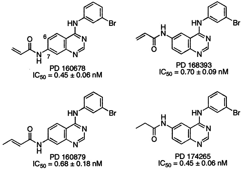
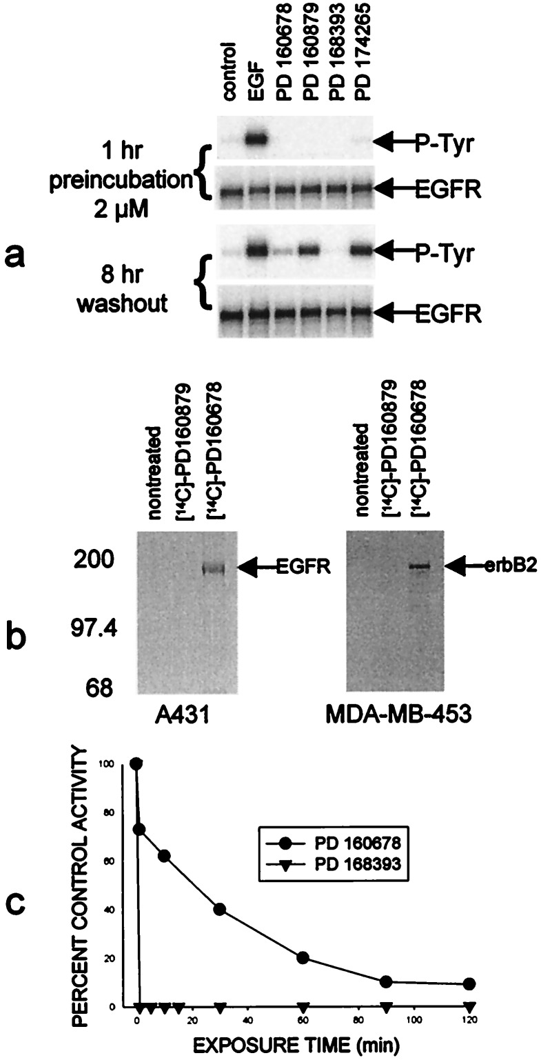
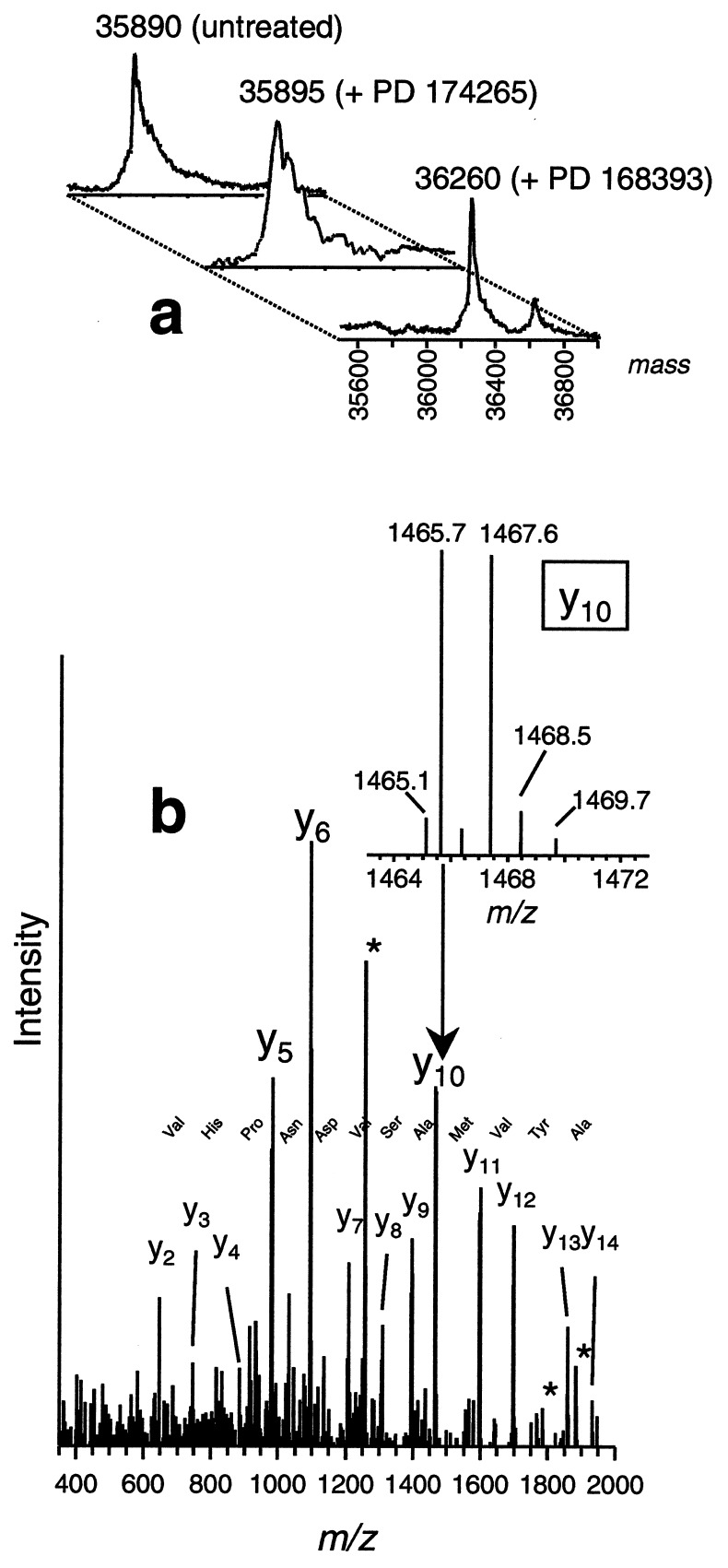
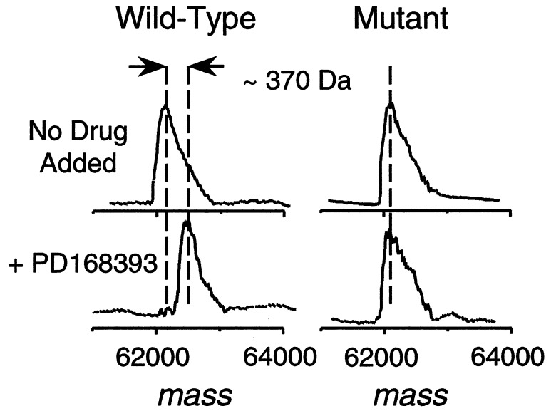
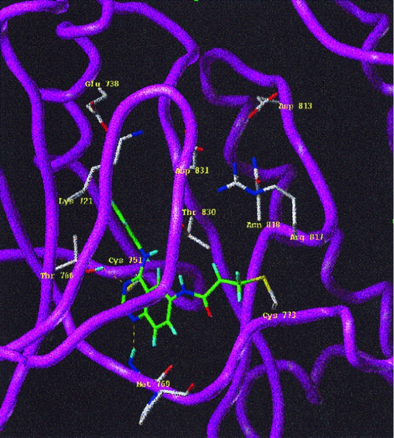
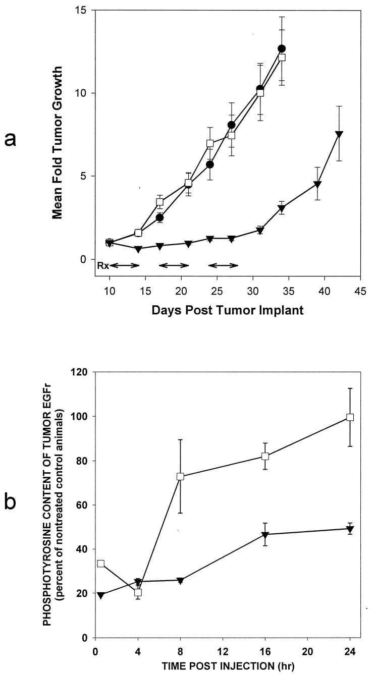
A class of high-affinity inhibitors is disclosed that selectively target and irreversibly inactivate the epidermal growth factor receptor tyrosine kinase through specific, covalent modification of a cysteine residue present in the ATP binding pocket. A series of experiments employing MS, molecular modeling, site-directed mutagenesis, and 14C-labeling studies in viable cells unequivocally demonstrate that these compounds selectively bind to the catalytic domain of the epidermal growth factor receptor with a 1:1 stoichiometry and alkylate Cys-773. While the compounds are essentially nonreactive in solution, they are subject to rapid nucleophilic attack by this particular amino acid when bound in the ATP pocket. The molecular orientation and positioning of the acrylamide group in these inhibitors in relation to Cys-773 entirely support these results as determined from docking experiments in a homology-built molecular model of the ATP site. Evidence is also presented to indicate that the compounds interact in an analogous fashion with erbB2 but have no activity against the other receptor tyrosine kinases or intracellular tyrosine kinases that were tested in this study. Finally, a direct comparison between 6-acrylamido-4-anilinoquinazoline and an equally potent but reversible analog shows that the irreversible inhibitor has far superior in vivo antitumor activity in a human epidermoid carcinoma xenograft model with no overt toxicity at therapeutically active doses. The activity profile for this compound is prototypical of a generation of tyrosine kinase inhibitors with great promise for therapeutic significance in the treatment of proliferative disease.


Figures

Figure 1
Figure 1
Chemical structures for PD 160678, 160879, 168393, and 174265. The compounds were synthesized as described (35). The IC50 values represent the concentration of compound necessary to inhibit purified full-length EGFr TK activity by 50% ± SE and represent the average of three separate experiments.
Figure 2
Figure 2
(a) A431 human epidermoid carcinoma cells were grown in 6-well plates to ≈90% confluence and then incubated in serum-free media for 18 hr. Duplicate sets of cells were treated with 2 μM designated compound for 1 hr. One set of cells was then stimulated with 100 ng/ml EGF for 5 min, and extracts were made as described (33). The other set of cells was washed free of the compound with warmed compound-free medium, incubated for 2 hr, washed again, incubated another 2 hr, washed again, and then incubated a further 4 hr and then stimulated with EGF. (b) 14C-labeled PD 160678 permanently associates with EGFr and erbB2, but not the reversible congener, PD 160879. A431 human epidermoid carcinoma cells or MDA-MB-453 human breast carcinoma cells were exposed to 2 μM 14C-labeled PD 160678 or 160879 for 2 hr and excess compound was washed away. Extracts were made and either the EGFr or erbB2 was immunoprecipitated as described (33). (c) Placement of the acrylamide side chain at the 6 position of the quinazoline allows a much more rapid irreversible interaction than at the 7 position. Cells were pretreated with the designated compound for the times shown, washed as described for a, and assayed for autophosphorylation activity 8 hr later.
Figure 3
Figure 3
MS analysis of PD 168393 binding to EGFr TK or intracellular domain. (a) Deconvoluted ESI mass spectra of EGFr TK protein and protein after addition of PD 174265 or 168393, with the latter showing a mass increase of ≈370 Da, consistent for a 1:1 protein–drug covalent binding stoichiometry. A small amount (≈15% abundance) of a 1:2 protein-drug species (molecular mass 36,626 Da) is also observed in the spectrum. (b) Fragmentation mass spectrum (MS/MS) of the selected doubly charged tryptic peptide at mass-to-charge m/z 1266 produces predominantly C-terminal containing y-ion series ions, using the nomenclature of Biemann (42). Tandem mass spectra were acquired from an on-line liquid chromatography-MS/MS experiment. The continuous y-ion series and the few N-terminally containing b-ions observed (marked with *) are consistent with the tryptic fragment EILDEAYVMASVDNPHVCR, with the drug attached to the cysteine residue. (Inset) Blowup of the isotope peak pattern for Y10 indicating the nearly equal abundant M and M+2 peak pattern, consistent for a Br-containing ion resulting from addition of a Br-containing compound.
Figure 4
Figure 4
Deconvoluted ESI mass spectra of wild-type or mutant (C773S) EGFr intracellular domain as described in Fig. 3.
Figure 5
Figure 5
The reaction product of PD 168393 in the EGFr TK ATP pocket. The three-dimensional model was constructed by using the homology modeling module implemented in look (Molecular Applications Group, Palo Alto, CA ) with cAMP kinase as the template. The basis for the three-dimensional model was the sequence alignment recently published (43). PD 168393 was constructed in sybyl 6.2 (Trips E & S, St. Louis) and the charges were derived from the semi-empirical molecular orbital package mopac using the modified neglect of differential overlap (MNDO) approach (44). The inhibitor was docked manually into the ATP binding site and the geometry of the reaction product was derived by using structural information from the Cambridge crystallographic data bank (48). The geometry was then optimized with the Trips force field implemented in sybyl 6.2 to relieve unfavorable steric contacts.
Figure 6
Figure 6
Comparative antitumor activity of PD 168393 and its reversible congener PD 174265 (a) and corresponding modulation of the EGFr phosphotyrosine content (b). •, Control; □, PD 174265; ▾, PD 168393. (a) Athymic nude mice housed in filtered cages were implanted s.c. with a fragment (≈30 mg) of A431 human epidermoid carcinoma and were randomized into treatment groups when the tumors were palpable. Animal dosing and tumor measurement were carried out essentially as described (45). The compounds were suspended in a vehicle containing 4% dimethylacetamide in aqueous 50 mM sodium lactate buffer (pH 4) and delivered i.p. to the mice at 58 mg/kg on days 10–14, 17–21, and 24–28 (shown with arrows) after tumor implant. Bars = mean ± SE. (b) Nude mice bearing 300- to 500-mg A431 tumors were treated with a single 58 mg/kg i.p. dose of either inhibitor or vehicle as described above. At various times after treatment the tumors were excised and ground to a frozen powder with a mortar and pestle chilled with liquid nitrogen. EGFr was extracted (33) and phosphotyrosine content was determined by ELISA (Calbiochem).

References

    1. Carraway K L, Cantley L C. Cell. 1994;78:5–8. - PubMed
    1. Pinkas-Kramarski R, Soussan L, Waterman H, Levkowitz G, Alroy I, Klapper L, Lavi S, Seger R, Ratzkin B J, Sela M, Yarden Y. EMBO J. 1996;15:2452–2467. - PMC - PubMed
    1. Reese D M, Slamon D J. Stem Cells. 1997;15:1–8. - PubMed
    1. Burke C L, Lemmon M A, Coren B A, Engelman D M, Stern D F. Oncogene. 1997;14:687–696. - PubMed
    1. Graus-Porta D, Beerli R R, Daly J M, Hynes N E. EMBO J. 1997;16:1647–1655. - PMC - PubMed

MeSH terms

LinkOut - more resources