

Neuroimaging analyses of human working memory
- PMID: 9751790
- PMCID: PMC21765
- DOI: 10.1073/pnas.95.20.12061
Neuroimaging analyses of human working memory
Abstract
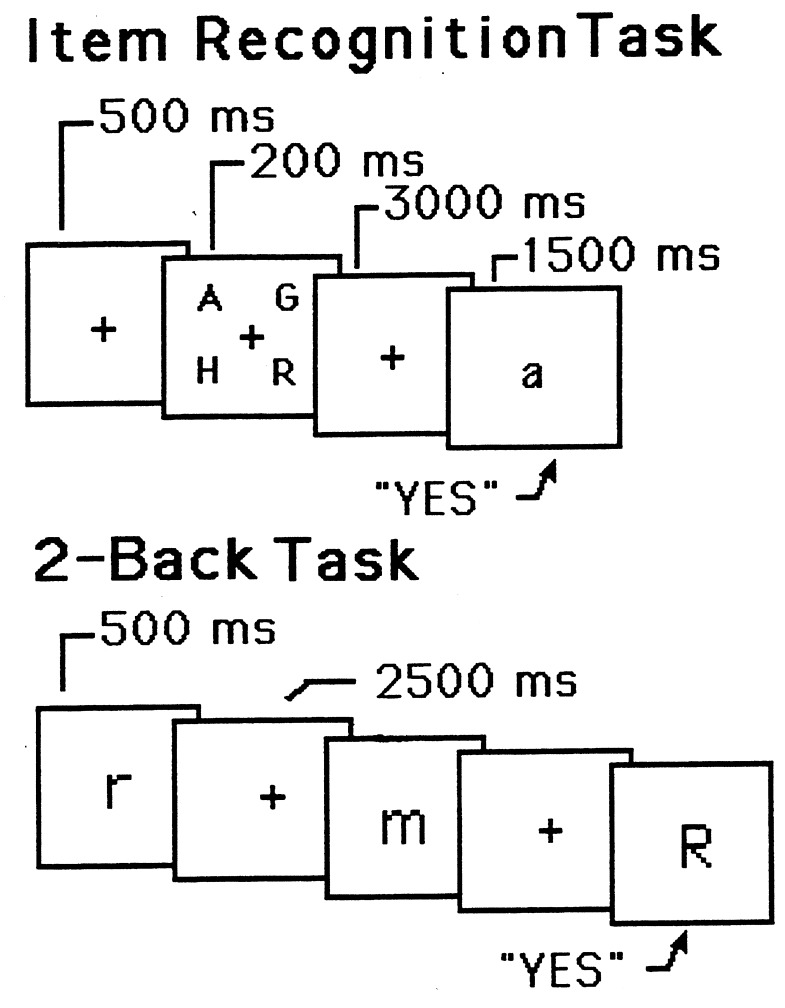
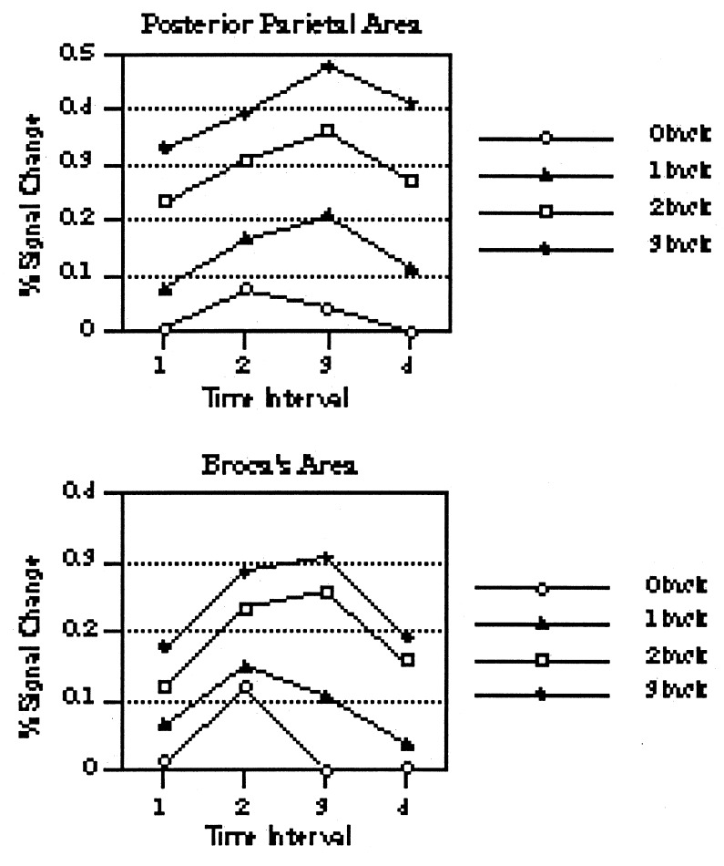
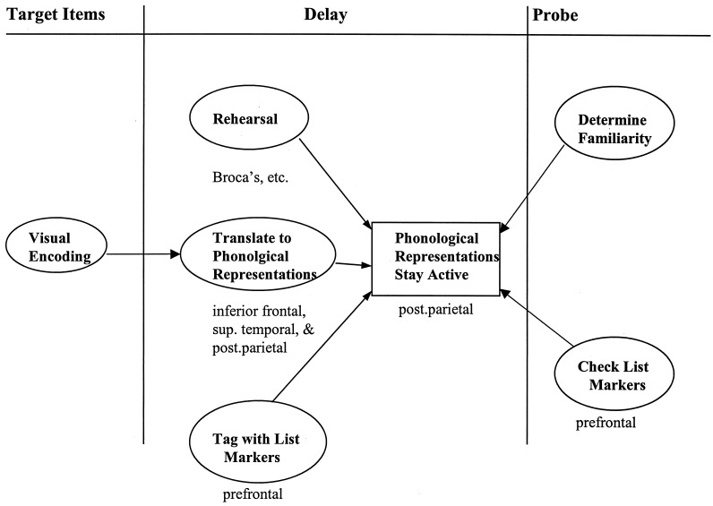
We review a program of research that uses neuroimaging techniques to determine the functional and neural architecture of human working memory. A first set of studies indicates that verbal working memory includes a storage component, which is implemented neurally by areas in the left-hemisphere posterior parietal cortex, and a subvocal rehearsal component, which is implemented by left-hemisphere speech areas, including Broca's area as well as the premotor and supplementary motor areas. We provide a number of neuroimaging dissociations between the storage and rehearsal areas. A second set of studies focuses on spatial working memory and indicates that it is mediated by a network of predominantly right-hemisphere regions that include areas in posterior parietal, occipital, and frontal cortex. We provide some suggestive evidence that these areas, too, divide into storage and rehearsal regions, with right-hemisphere posterior parietal and premotor regions subserving spatial rehearsal. In a final set of studies, we turn to "executive processes," metaprocesses that regulate the processing of working-memory contents. We focus on the executive process of inhibition as it is used in verbal working memory. We provide evidence that such inhibition is mediated by the left-hemisphere prefrontal region and that it can be dissociated from verbal storage and rehearsal processes.
Figures

References
-
- Baddeley A D. Science. 1992;225:556–559. - PubMed
-
- Carpenter P A, Just M A, Shell P. Psychol Rev. 1990;97:404–431. - PubMed
-
- Jonides J. In: An Invitation to Cognitive Science: Thinking. Smith E E, Osherson D, editors. Vol. 3. Cambridge, MA: MIT Press; 1995. pp. 215–265.
-
- Jonides J, Reuter-Lorenz P, Smith E E, Awh E, Barnes L, Drain M, Glass J, Lauber E, Patalano A, Schumacher E H. In: The Psychology of Learning and Motivation. Medin D, editor. New York: Academic; 1996. pp. 43–88.
-
- Shulman G L, Fiez J A, Corbetta M, Buckner R L, Miezin F M, Raichle M E, Petersen S E. J Cognit Neurosci. 1997;9:648–663. - PubMed
Publication types
MeSH terms
LinkOut - more resources
Full Text Sources
Medical
