

Management of breast fibroadenomas
- PMID: 9754521
- PMCID: PMC1497021
- DOI: 10.1046/j.1525-1497.1998.cr188.x
Management of breast fibroadenomas
Abstract
Objective: To identify from the literature and clinical experience a rational approach to management of fibroadenomas of the breast.
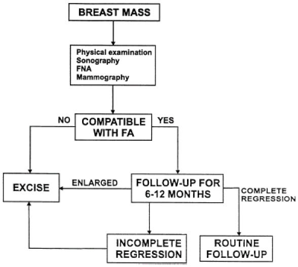
Method: Recent literature on detection, diagnosis, and natural history of fibroadenomas was reviewed. Experience with over 4,000 women evaluated in the breast clinic at the Tel-Aviv Medical Center contributed to the management strategies suggested by review of the literature.
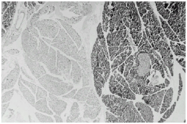
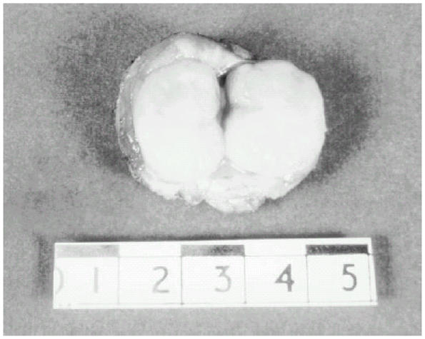
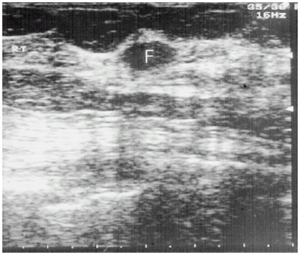
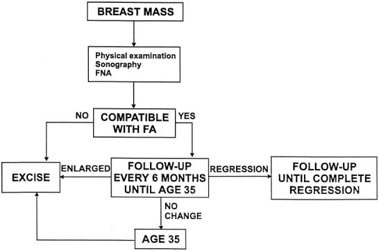
Results: Fibroadenomas of the breast are common, accounting for 50% of all breast biopsies performed. Physical examination, sonography, and fine needle aspiration are effective in distinguishing fibroadenomas from breast cancer. Transformation from fibroadenoma to cancer is rare; regression or resolution is frequent, supporting conservative approaches to follow-up and management.
Conclusion: Age-based algorithms that allow for conservative management and that limit excision to patients whose fibroadenomas fail to regress are presented.
Figures
References
-
- Dent DM, Hacking EA, Wilkie W. Benign breast disease clinical classification and disease distribution. Br J Clin Pract. 1988;42(suppl 56):69–71.
-
- Franyz VK, Pickern JW, Melcher GW, Auchincoloss JR. Incidence of chronic cystic disease in so-called normal breast: a study based on 225 post mortem examinations. Cancer. 1951;4:762–7. - PubMed
-
- Schuerch C, Rosen PP, Hirota T, Itabashi M. A pathologic study of benign breast disease in Tokyo and New York. Cancer. 1982;50:1899–902. - PubMed
-
- Onuigb WIB. Adolescent mass in Nigerian igbos. Am J Surg. 1979;137:367–71. - PubMed
-
- Brinton LA, Vajsey MP, Flavel R. Risk factors for benign breast disease. Am J Epidemiol. 1981;113:203–14. - PubMed
Publication types
MeSH terms
LinkOut - more resources
Full Text Sources
Medical