. 1998 Oct;42(10):2467-73.
doi: 10.1128/AAC.42.10.2467.

Granulocyte colony-stimulating factor and azole antifungal therapy in murine aspergillosis: role of immune suppression

Affiliations

Granulocyte colony-stimulating factor and azole antifungal therapy in murine aspergillosis: role of immune suppression

J R Graybill et al. Antimicrob Agents Chemother. 1998 Oct.

Abstract

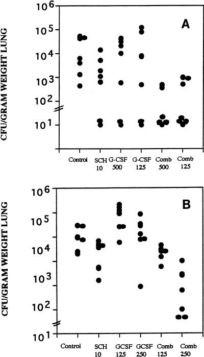
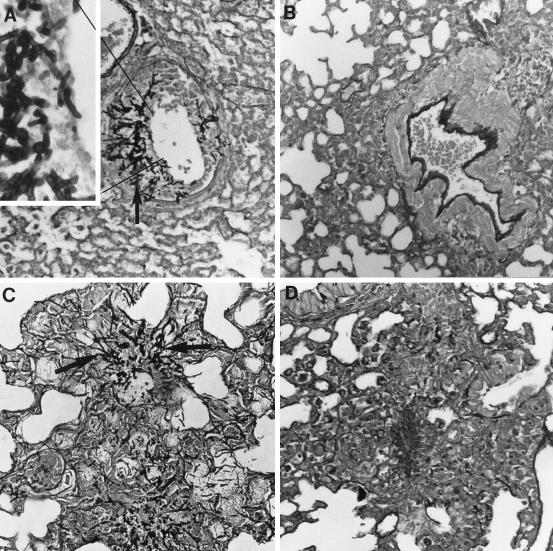
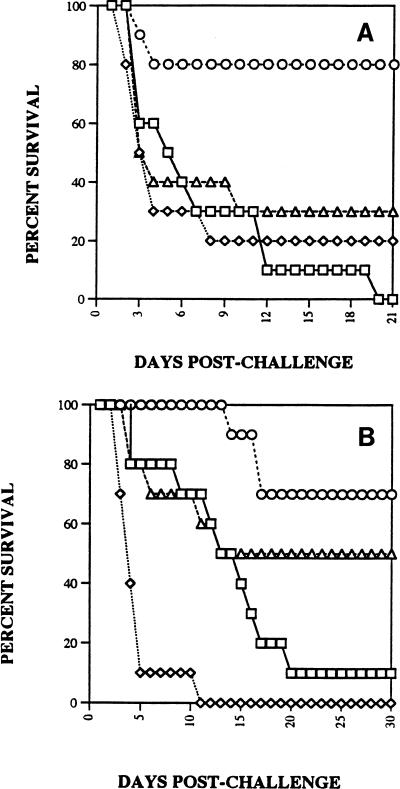
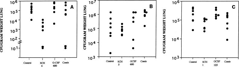
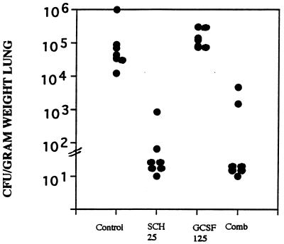
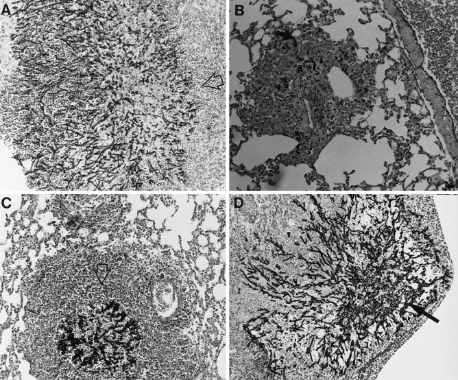
Outbred ICR mice were immune suppressed either with hydrocortisone or with 5-fluorouracil and were infected intranasally with Aspergillus fumigatus. Beginning 3 days before infection some groups of mice were given recombinant human granulocyte colony-stimulating factor (G-CSF), SCH56592 (an antifungal triazole), or both. Corticosteroid-pretreated mice responded to SCH56592 and had reduced counts in lung tissue and prolonged survival. In these mice, G-CSF strongly antagonized the antifungal activity of SCH56592. Animals treated with both agents developed large lung abscesses with polymorphonuclear leukocytes and large amounts of Aspergillus. In contrast, mice made neutropenic with 5-fluorouracil and then infected with A. fumigatus conidia benefited from either G-CSF or triazoles, and the effect of the combination was additive rather than antagonistic. Host predisposing factors contribute in different ways to the outcome of growth factor therapy in aspergillosis.


Figures

FIG. 1
FIG. 1
Burden of A. fumigatus in lung tissue. Groups of seven mice each were treated with G-CSF beginning on day −3 before infection, with cyclophosphamide at 200 mg/kg and 5-fluorouracil at 150 mg/kg on the day of infection, and with SCH56592 beginning 1 day after infection. Treatment was stopped on day 5 after infection, and mice were killed on day 6 after infection. (A) Mice were infected with >108 conidia, and only combined (Comb) treatment with SCH56592 at 10 mg/kg/dose and GCSF at 125 or 500 μg/kg significantly reduced tissue burden below that for the untreated controls. (B) Mice were infected with 3.7 × 10⁷ CFU, and the effects of G-CSF at 125 or 250 μg/kg alone, SCH56592 alone, and the combined (Comb) treatment regimen of SCH56592 and G-CSF were evaluated; only the combined treatment regimen with G-CSF at 250 μg/kg/day significantly lowered the lung tissue counts below that for the untreated controls.
FIG. 2
FIG. 2
Histopathology of lungs of neutropenic mice infected with 107 CFU conidia, treated from days 1 through 6, and killed. There were three mice per group. Representative sections are shown at × 440 magnification, with the inset at × 1,000 magnification. The stain was hematoxylin and eosin combined with Gomori’s methenamine silver. (A) Untreated control mice. Two mice had extensive mycotic bronchitis and one had minimal infiltrates, which were monocytic. Large numbers of hyphae were seen invading the bronchioles in two mice (the inset shows hyphae invading a bronchiole; the solid arrow indicates hyphae). (B) Mice treated with SCH56592 at 10 mg/kg/day. Minimal monocyte infiltrates were seen in two mice. No hyphae were seen. A lesion from one mouse is shown. (C) G-CSF at 600 μg/kg/day. Many large collections of PMNs were seen in one mouse, and mycotic bronchitis and pneumonia were observed in the others. Abundant hyphae were seen in lesions from all three mice (solid arrow). Some bacterial overgrowth is also apparent in this section. (D) G-CSF and SCH56592. Minimal mononuclear infiltrates were seen in all three mice, with some PMNs seen in the lesions of one mouse. No hyphae were seen. A lesion is shown.
FIG. 3
FIG. 3
Survival after intranasal infection of groups of 10 mice with conidia of A. fumigatus. Mice were treated with G-CSF at 600 μg/kg beginning on day −3 before infection and continuing through day 5 after infection. Hydrocortisone at 100 mg/kg was begun subcutaneously on day −2 before infection and was continued through day 1 after infection. SCH56592 was begun on day 1 and was continued through day 5 after infection. □, control; ◊, G-CSF at 600 μg/kg; ○, SCH56592; ▵, G-CSF at 600 μg/kg and SCH56592. (A) Mice were infected with 1.3 × 10⁸ CFU. Only SCH56592 at 5 mg/kg/day significantly prolonged survival beyond that for untreated controls. (B) Mice were infected with 2.2 × 10⁶ CFU. SCH56592 at 25 mg/kg/day alone prolonged survival. The response to combined therapy was significantly shorter than that to therapy with SCH56592 alone.
FIG. 4
FIG. 4
Lung tissue burden of A. fumigatus as CFU per gram of lung weight. (A) Mice were concurrently infected with those in the survival study (Fig. 3A) and were infected with 1.3 × 10⁸ CFU. Only SCH56592 at 5 mg/kg/day (SCH 5) significantly reduced the tissue burden. Slashed bars across the y axis indicate the minimum detectable counts, usually 20 to 30 CFU/g of lung tissue. (B) Mice were infected with 4 × 10⁷ CFU and were given G-CSF at 600 mg/kg/day beginning on day −3. The G-CSF and combined (Comb) treatment groups had only seven mice because of cannibalism of several mice that succumbed early. No significant differences were seen, although the result for SCH56592 alone was close to significance compared with the result for the controls. (C) Mice were infected with 107 CFU and were given either SCH56592 at a low dose of 1 mg/kg/day (SCH 1), GCSF at 125 μg/kg/day, or both. No significant reduction in counts in tissue compared with the counts for untreated controls was noted for any group.
FIG. 5
FIG. 5
Lung tissue burden of A. fumigatus. Mice were infected with a sublethal inoculum of 4.5 × 10⁴ CFU and treated with SCH56592 at 25 mg/kg/dose, GCSF at 125 μg/kg/dose, or both (Comb). With this regimen SCH56592 alone and combined therapy reduced the counts in lung tissue significantly more than no treatment did. Combined therapy was not superior to SCH56592 alone.
FIG. 6
FIG. 6
Histopathology of the lungs of mice 6 days after infection with 107 A. fumigatus. Hematoxylin and eosin stain combined with Gomori’s methenamine silver was used. Three mice were examined for each treatment group. Magnifications, × 400. (A) Untreated control mice. Large numbers of hyphae are seen invading the bronchioles. Foci of hyphae are surrounded by a modest infiltrate of predominantly monocytic cells with a few PMNs (open arrow). (B) Mice treated with SCH56592 at 10 mg/kg/day. Few hyphae are seen (solid arrow), with modest surrounding infiltrates of mononuclear cells. (C) G-CSF at 600 μg/kg/day. Many large collections of PMNs and necrotic debris (open arrow) surround foci with large numbers of hyphae present. (D) G-CSF and SCH56592. Large abscesses with PMNs surrounding foci of A. fumigatus hyphae. Hyphae (solid arrow) are more numerous than in mice treated with SCH56592 alone.

References

    1. Adelman B A, Bentman A, Rosenthal P, Smith B R, Bridges K R, Holmes W. Treatment of aspergillosis in leukemia. Ann Intern Med. 1979;91:323–324. - PubMed
    1. Albelda S M, Talbot G H, Gerson S L, Miller W T, Cassileth P A. Pulmonary cavitation and massive hemoptysis in invasive pulmonary aspergillosis: influence of bone marrow recovery in patients with acute leukemia. Am Rev Respir Dis. 1985;131:115–120. - PubMed
    1. Andriole, V. T. 1993. Infections with Aspergillus species. Clin. Infect. Dis. 17(Suppl. 2):S481–S486. - PubMed
    1. Berenguer J, Allende M C, Lee J W, Garret K, Lyman C, Ali N M, Bacher J, Pizzo P A, Walsh T J. Pathogenesis of pulmonary aspergillosis—granulocytopenia versus cyclosporine and methylprednisolone-induced immunosuppression. Am J Respir Crit Care Med. 1995;152:1079–1086. - PubMed
    1. Binder R E, Faling L J, Pugatch R D, Mahasaen C, Snider G L. Chronic necrotizing pulmonary aspergillosis: a discrete clinical entity. Medicine (Baltimore) 1982;61:109–124. - PubMed

Publication types