. 1998 Oct;42(10):2731-8.
doi: 10.1128/AAC.42.10.2731.

Proteasome inhibitors block development of Plasmodium spp

Affiliations

Proteasome inhibitors block development of Plasmodium spp

S M Gantt et al. Antimicrob Agents Chemother. 1998 Oct.

Abstract

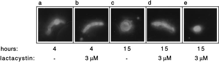
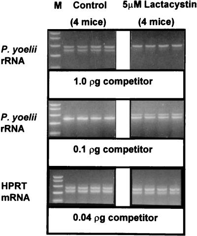
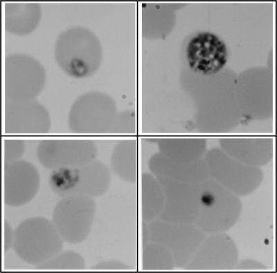
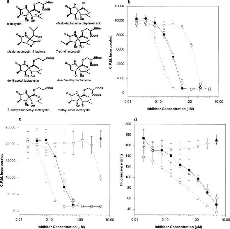
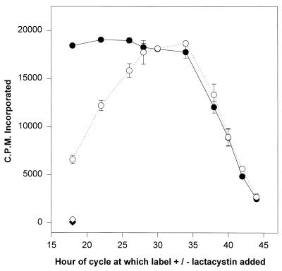
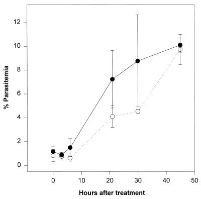
Proteasomes degrade most of the proteins inside eukaryotic cells, including transcription factors and regulators of cell cycle progression. Here we show that nanomolar concentrations of lactacystin, a specific irreversible inhibitor of the 20S proteasome, inhibit development of the exoerythrocytic and erythrocytic stages of the malaria parasite. Although lactacystin-treated Plasmodium berghei sporozoites are still invasive, their development into exoerythrocytic forms (EEF) is inhibited in vitro and in vivo. Erythrocytic schizogony of P. falciparum in vitro is also profoundly inhibited when drug treatment of the synchronized parasites is prior, but not subsequent, to the initiation of DNA synthesis, suggesting that the inhibitory effect of lactacystin is cell cycle specific. Lactacystin reduces P. berghei parasitemia in rats, but the therapeutic index is very low. Along with other studies showing that lactacystin inhibits stage-specific transformation in Trypanosoma and Entamoeba spp., these findings highlight the potential of proteasome inhibitors as drugs for the treatment of diseases caused by protozoan parasites.


Figures

FIG. 1
FIG. 1
Lactacystin alters the normal development of sporozoites into EEF in vitro. P. berghei sporozoites were incubated in 3 μM lactacystin or medium alone for 15 min and then added to HepG2 cells and allowed to invade and begin their development into EEF. Four and fifteen hours later the cells were fixed and stained with MAb 3D11 by using the double-staining assay, which allows the distinction between intracellular and extracellular sporozoites. Magnification, ×100.
FIG. 2
FIG. 2
Lactacystin inhibits the switch to A-type rRNA of P. berghei in vitro. Sporozoites were incubated with 3 μM lactacystin or without lactacystin for 15 min and then plated on HepG2 cells. After 2 h, the medium was removed and fresh medium without inhibitor was added. At 5 (a) and 21 h (b), total RNA was extracted and RT reactions were performed with 0.1 μg of RNA. Quantitative PCR of the cDNA was performed with primers specific for A-type rRNA and serial dilutions of cDNA. The first, second, and third (panel b only) lanes in each panel show the results of PCR performed with 2, 0.4, and 0.2 μl, respectively, of cDNA.
FIG. 3
FIG. 3
Lactacystin decreases sporozoite infectivity in vivo. P. yoelii sporozoites were incubated in 5 μM lactacystin or medium alone for 1 h. Two thousand sporozoites were then injected i.v. into each mouse, and 40 h later the mouse livers were harvested for isolation of RNA. Sporozoite infectivity was quantified by measuring the amount of parasite rRNA in a quantitative RT-PCR assay. The top two panels show PCRs performed with P. yoelii rRNA primers and 1 and 0.1 pg of a P. yoelii rRNA competitor. The parasite target band is 393 bp, and that of the competitor is 459 bp. The bottom panel shows control PCRs performed with the same RT reaction mixtures containing HPRT primers and 0.04 pg of an HPRT competitor; the HPRT target band is 352 bp, and that of the competitor is 450 bp. M = markers (1,000, 750, 500, 300, and 150 bp).
FIG. 4
FIG. 4
Lactacystin inhibits the development of P. falciparum erythrocytic stages in vitro. Photomicrographs were taken of synchronized trophozoites at 18 h (top left), after which the trophozoites were incubated for another 24 h in either medium alone (top right) or 1.25 (bottom left) or 10 μM (bottom right) lactacystin.
FIG. 5
FIG. 5
Lactacystin analogs inhibit P. falciparum erythrocytic stages and isolated human proteasomes similarly. (a) Chemical structures of lactacystin and the analogs studied. (b and c) Synchronized trophozoites at 18 h of the erythrocytic cycle were plated in 96-well microtiter plates with [3H]hypoxanthine and the concentrations of inhibitors indicated. After 24 h, cells were harvested and incorporation of the label was measured. Shown are the means of triplicate wells ± standard deviations. (d) Proteasomes isolated from normal human erythrocytes, plus inhibitors at the indicated concentrations, were incubated with fluorogenic substrate N-succinyl-Leu-Leu-Val-Tyr-7-amido-4-methyl-coumarin to measure chymotrypsin-like activity. Each point is the mean of the fluorescences of duplicate wells ± the standard deviation. Symbols (panels b, c, and d): solid circles, lactacystin; open circles, clasto-lactacystin β-lactone; open upright triangles, β-acetylaminoethyl lactacystin; open diamonds, de-N-acetyl lactacystin; solid triangles, clasto-lactacystin dihydroxy acid; open hexagons, 7-ethyl lactacystin; open inverted triangles, des-7-methyl lactacystin; open squares, methyl ester lactacystin.
FIG. 6
FIG. 6
Lactacystin inhibits DNA synthesis in a stage-specific manner. Synchronized trophozoites were plated in 96-well microtiter plates at 18 h into the erythrocytic cycle. [3H]hypoxanthine with (open circles) or without (closed circles) lactacystin was added to wells at the times indicated. All cells were harvested at 48 h in the cycle, and incorporation of the label was measured. Shown are the means of triplicate wells with standard deviations.
FIG. 7
FIG. 7
Lactacystin significantly reduces parasitemia in vivo. Six P. berghei-infected rats were paired into two groups of three rats; all rats had comparable parasitemias. Each rat in the experimental group (open circles) received 1.6 mg of lactacystin in 1 ml of PBS, given as one intraperitoneal injection of 0.5 ml and one i.v. injection of 0.5 ml at the same time. Each rat in the control group (solid circles) received identical injections of PBS alone. Giemsa-stained blood smears, taken at the time points indicated, were blindly counted to measure parasitemias. Each point represents the mean of parasitemias from three rats ± the standard deviation.

References

    1. Aikawa M, Schwartz A, Uni S, Nussenzweig R, Hollingdale M. Ultrastructure of in vitro cultured exoerythrocytic stage of Plasmodium berghei in a hepatoma cell line. Am J Trop Med Hyg. 1984;33:792–799. - PubMed
    1. Briones M R S, Tsuji M, Nussenzweig V. The large differences in infectivity for mice of Plasmodium berghei and Plasmodium yoelii sporozoites cannot be correlated with their ability to enter hepatocytes. Mol Biochem Parasitol. 1996;77:7–17. - PubMed
    1. Butler D, Maurice J, O’Brien C. Time to put malaria control on the global agenda. Nature. 1997;386:535–540. - PubMed
    1. Corey E J, Reichard G A. Total synthesis of lactacystin. J Am Chem Soc. 1992;114:10677–10678.
    1. Coux O, Tanaka K, Goldberg A L. Structure and functions of the 20S and 26S proteasomes. Annu Rev Biochem. 1996;65:801–847. - PubMed

Publication types

MeSH terms

LinkOut - more resources