. 1998 Oct;64(10):3656-62.
doi: 10.1128/AEM.64.10.3656-3662.1998.

A bioluminescence assay using Nitrosomonas europaea for rapid and sensitive detection of nitrification inhibitors

Affiliations

A bioluminescence assay using Nitrosomonas europaea for rapid and sensitive detection of nitrification inhibitors

T Iizumi et al. Appl Environ Microbiol. 1998 Oct.

Abstract

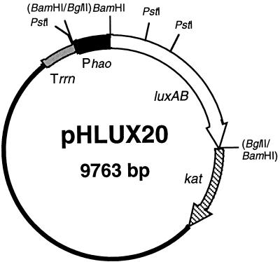
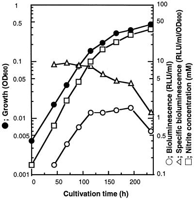
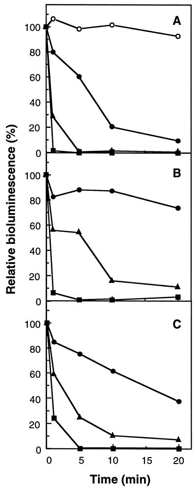
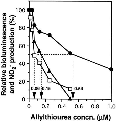
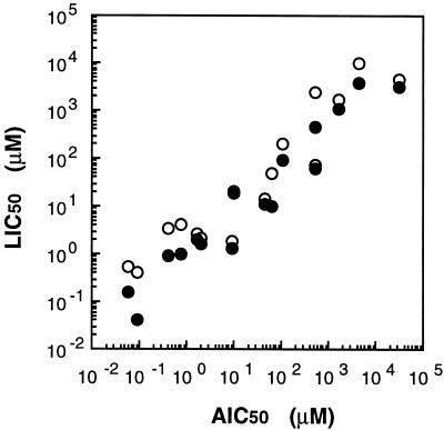
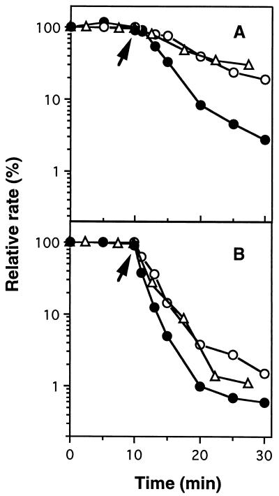
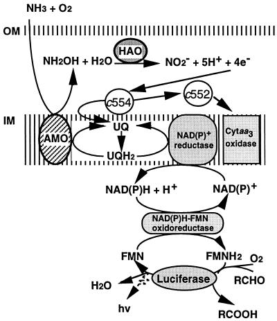
An expression vector for the luxAB genes, derived from Vibrio harveyi, was introduced into Nitrosomonas europaea. Although the recombinant strain produced bioluminescence due to the expression of the luxAB genes under normal growing conditions, the intensity of the light emission decreased immediately, in a time-and dose-dependent manner, with the addition of ammonia monooxygenase inhibitors, such as allylthiourea, phenol, and nitrapyrin. When whole cells were challenged with several nitrification inhibitors and toxic compounds, a close relationship was found between the change in the intensity of the light emission and the level of ammonia-oxidizing activity. The response of bioluminescence to the addition of allylthiourea was considerably faster than the change in the ammonia-oxidizing rate, measured as both the O2 uptake and NO2- production rates. The bioluminescence of cells inactivated by ammonia monooxygenase inhibitor was recovered rapidly by the addition of certain substrates for hydroxylamine oxidoreductase. These results suggested that the inhibition of bioluminescence was caused by the immediate decrease of reducing power in the cell due to the inactivation of ammonia monooxygenase, as well as by the destruction of other cellular metabolic pathways. We conclude that the assay system using luminous Nitrosomonas can be applied as a rapid and sensitive detection test for nitrification inhibitors, and it will be used to monitor the nitrification process in wastewater treatment plants.


Figures

FIG. 1
FIG. 1
Physical map of pHLUX20. Promoterless luciferase-encoding genes (luxAB) from V. harveyi and the Tn903-derived kanamycin acetyltransferase-encoding gene (kat) are shown as open and striped arrows, respectively, indicating the gene orientations. The E. coli 5S rRNA rho-independent terminator (Trrn) and the promoter region of the N. europaea HAO-encoding gene (Phao) are represented by shaded and solid bars, respectively. The solid line is the region derived from the IncQ plasmid, which is essential for replication.
FIG. 2
FIG. 2
Results of cultivation of N. europaea(pHLUX20). N. europaea(pHLUX20) was grown in P medium by using a 5-liter jar fermentor with a working volume of 3.5 liters at 30°C. Details of cultivation conditions are given in Materials and Methods. Cell growth was monitored at 600 nm with cuvettes of a 50-mm light path. Bioluminescence was measured by a model 20e luminometer (Turner Design Co.) as described in Materials and Methods. The nitrite concentration of the culture supernatant was measured by colorimetric assay (7).
FIG. 3
FIG. 3
Effects of AMO inhibitors on bioluminescence in N. europaea(pHLUX20). An aliquot (0.95 ml) of the culture broth of N. europaea(pHLUX20) was placed in a test tube, and 50 μl of test sample was added. After incubation for 1 to 20 min at 25°C, the bioluminescence of the incubation mixture was measured. The bioluminescence of the control reaction just after the addition of water instead of inhibitor was 1.02 RLU/ml and is defined as 100% relative bioluminescence. Values are averages from three independent experiments. (A) Effect of allylthiourea. ○, control (H2O addition); •, 0.1 μM; ▴, 1 μM; ■, 10 μM. (B) Effect of phenol. •, 1 μM; ▴, 10 μM; ■, 100 μM. (C) Effect of nitrapyrin. •, 0.1 μM; ▴, 1 μM; ■, 10 μM. Inhibitor concentrations given are final concentrations in the incubation mixture.
FIG. 4
FIG. 4
Dose-response curve of allylthiourea and determination of the LIC50s and AIC50s. The strength of inhibition of light emission in N. europaea (pHLUX20) was expressed as the LIC50, which was calculated from the graphed data as 0.54 and 0.15 μM when the reaction mixture was incubated for 1 (•) and 5 (▴) min, respectively. On the other hand, the strength of inhibition of ammonia-oxidizing activity in N. europaea was expressed as the AIC50, which was calculated as 0.06 μM for the 30-min incubation (□). Values are averages from three independent experiments.
FIG. 5
FIG. 5
Correlation between LIC50s and AIC50s. Sixteen different nitrification inhibitors and toxic compounds were used. Data were plotted as shown in Table 1. LIC50s were determined by incubations of 1 (○) and 5 (•) min.
FIG. 6
FIG. 6
Comparison of bioluminescence and the ammonia-oxidizing rate in the presence of allylthiourea. The intensity of bioluminescence (•) and the ammonia-oxidizing rate, which was measured as both the O2 uptake rate (○) and the NO2 production rate (▵), were expressed as relative ratios. An N. europaea(pHLUX20) cell suspension (2.0 mg of protein/ml) of 500 μl was injected into the DO electrode-mounted flask containing 64 ml of 100 mM phosphate buffer (pH 7.8) with 19 mM (NH4)2SO4 (time zero) and was preincubated for 10 min with agitation by using a stirrer magnet at 25°C to establish the steady-state O2 uptake rate. Allylthiourea was then added at final concentrations of 0.1 (A) and 0.5 (B) μM after preincubation (indicated by arrows), and subsequent incubation was continued under the same conditions. The initial intensity of bioluminescence was 1,090 RLU/mg of protein. Steady-state rates of O2 uptake and NO2 production were 0.71 and 0.43 μmol/min/mg of protein, respectively. Values are averages from at least two independent experiments.
FIG. 7
FIG. 7
Hypothetical model of interaction between the electron transfer pathways and the luciferase reaction in N. europaea. The nitrification pathway is based on that proposed by Wood (30) and Hooper et al. (10). NAD(P)+ reductase and NAD(P)H-FMN oxidoreductase have not been identified in N. europaea. HAO generates reducing power by the oxidation of hydroxylamine, which is produced by the preceding oxidation of ammonia by AMO. The reducing power is then transferred to ubiquinone (UQ) via cytochrome c-554 (c554), and the resulting ubiquinol (UQH2) is thought to supply reducing power for the maintenance of the AMO reaction and the reduction of NAD(P)+. The remaining electrons from cytochrome c-554 may pass through cytochrome c-552 (c552) to cytochrome aa3 oxidase (Cytaa3 oxidase). NAD(P)+ is reduced by reverse electron transfer, which needs energy supplied by the hydrolysis of ATP. The reducing power for the luciferase reaction must be obtained from FMNH2, which is generated from the reduction of FMN in the NAD(P)H-FMN oxidoreductase reaction by using the reducing power of NAD(P)H. OM and IM, outer and inner membranes, respectively.

References

    1. Aleem M I H. Generation of reducing power in chemosynthesis. II. Energy linked reduction of pyridine nucleotide in the chemoautotroph Nitrosomonas europaea. Biochim Biophys Acta. 1966;113:216–224. - PubMed
    1. Bédard C, Knowles R. Physiology, biochemistry, and specific inhibitors of CH4, NH4+, and CO oxidation by methanotrophs and nitrifiers. Microbiol Rev. 1989;53:68–84. - PMC - PubMed
    1. Bergmann D J, Arciero D M, Hooper A B. Organization of the hao gene cluster of Nitrosomonas europaea: genes for two tetraheme c cytochromes. J Bacteriol. 1994;176:3148–3153. - PMC - PubMed
    1. Berks B C, Ferguson S J, Moir J W B, Richardson D J. Enzyme and associated electron transport systems that catalyse the respiratory reduction of nitrogen oxides and oxyanions. Biochim Biophys Acta. 1995;1232:97–173. - PubMed
    1. Bulich A A, Greene M W, Isenberg D L. A practical and reliable method for monitoring the toxicity of aquatic samples. Process Biochem. 1982;17:45–47.

MeSH terms

LinkOut - more resources