. 1998 Oct 5;143(1):13-22.
doi: 10.1083/jcb.143.1.13.

Large-scale chromosomal movements during interphase progression in Drosophila

Affiliations

Large-scale chromosomal movements during interphase progression in Drosophila

A K Csink et al. J Cell Biol. .

Abstract

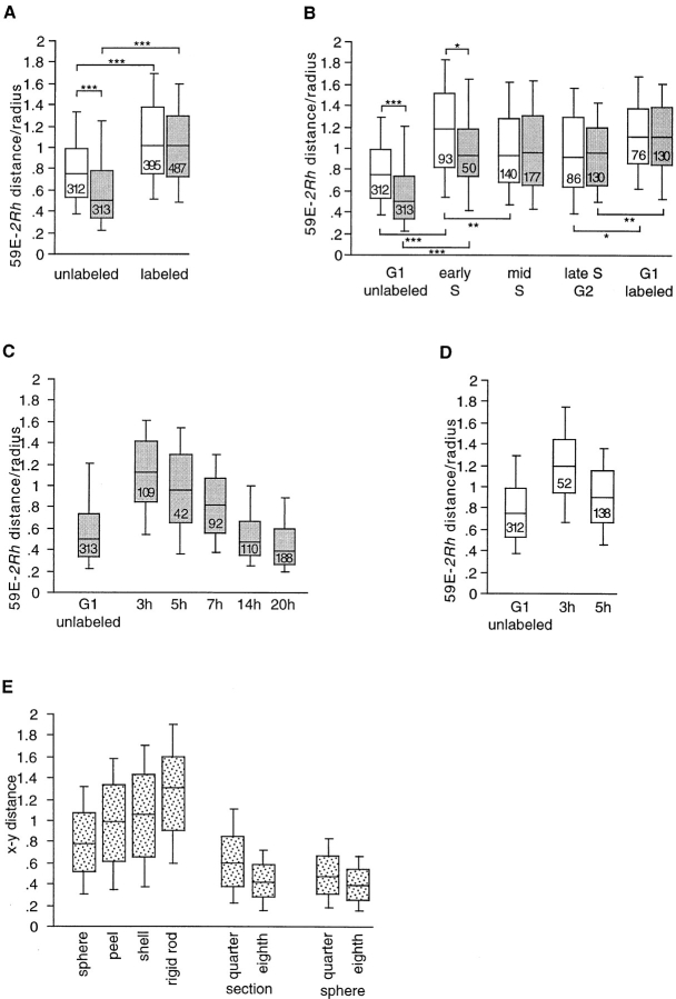
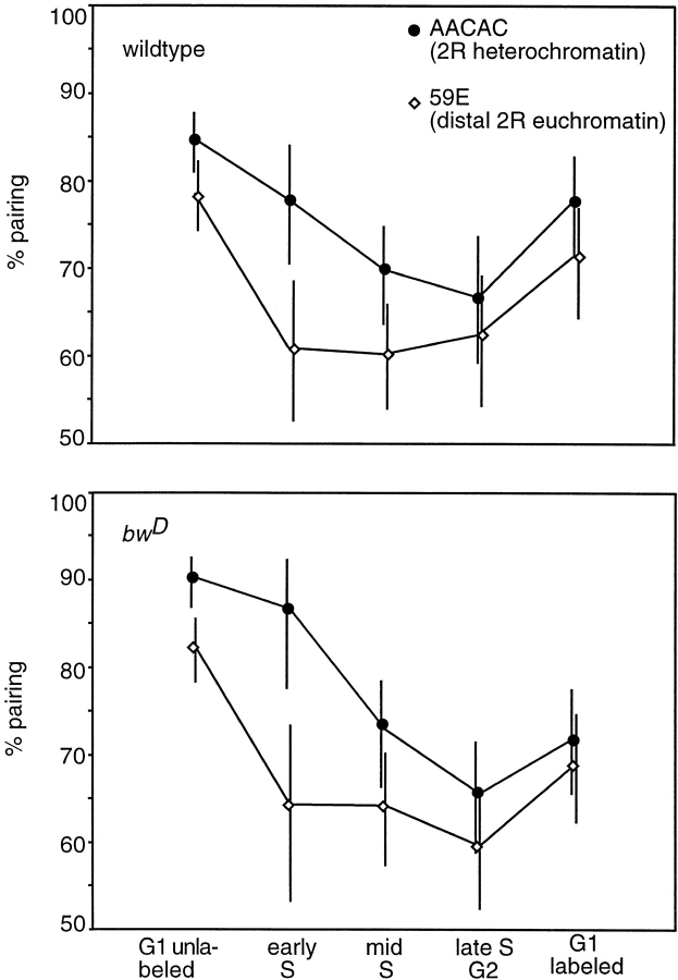
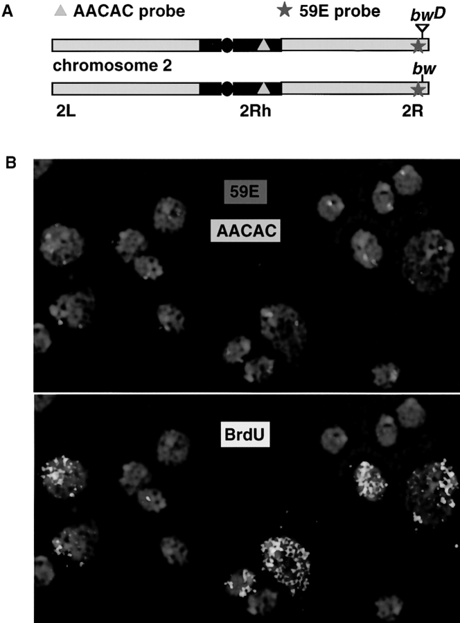
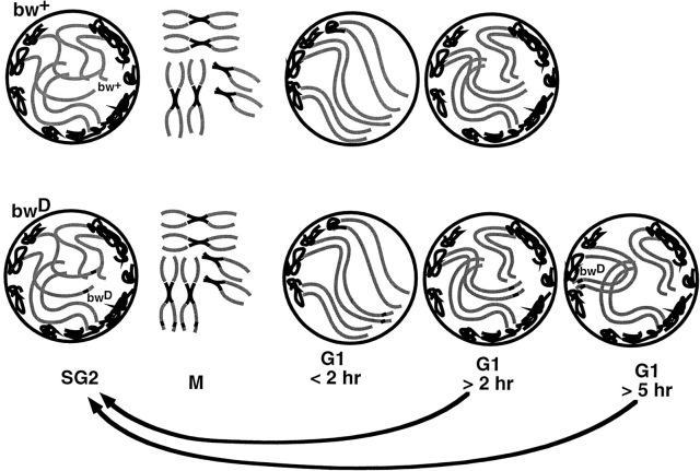
We examined the effect of cell cycle progression on various levels of chromosome organization in Drosophila. Using bromodeoxyuridine incorporation and DNA quantitation in combination with fluorescence in situ hybridization, we detected gross chromosomal movements in diploid interphase nuclei of larvae. At the onset of S-phase, an increased separation was seen between proximal and distal positions of a long chromsome arm. Progression through S-phase disrupted heterochromatic associations that have been correlated with gene silencing. Additionally, we have found that large-scale G1 nuclear architecture is continually dynamic. Nuclei display a Rabl configuration for only approximately 2 h after mitosis, and with further progression of G1-phase can establish heterochromatic interactions between distal and proximal parts of the chromosome arm. We also find evidence that somatic pairing of homologous chromosomes is disrupted during S-phase more rapidly for a euchromatic than for a heterochromatic region. Such interphase chromosome movements suggest a possible mechanism that links gene regulation via nuclear positioning to the cell cycle: delayed maturation of heterochromatin during G1-phase delays establishment of a silent chromatin state.


Figures

Figure 1
Figure 1
Distribution of BrdU labeling patterns and DNA content in larval CNS nuclei during the cell cycle. Box plots of the relative DAPI intensity, wherein each horizontal line represents the 10th, 25th, 50th (median), 75th and 90th percentiles. The numbers in the boxes are the total number of nuclei in each plot. Larvae were fed BrdU for three different time periods, and the nuclei were analyzed for BrdU labeling pattern. A euchromatic pattern consists of fine-grained labeling over most of the nucleus, while a heterochromatic pattern consists of labeling in only a few bright spots. Undetermined nuclei were either entirely labeled or had a pattern that was not identifiable as heterochromatic or euchromatic.
Figure 3
Figure 3
Cell cycle–dependent chromosome movements. Box plots of the distance between the 2Rh probe (AACAC) and the 59E probe divided by the radius of the nucleus. Data from wild-type nuclei are shown in white boxes, and bwD nuclei are shown in gray boxes. Simulated distributions are shown in spotted boxes. The numbers in the boxes are the total number of measurements in each plot. The asterisks on the brackets indicated the level of significance for the bracketed pair as determined using the two-tailed Mann-Whitney U test for nonparametric comparison of two unpaired groups. *P < 0.05; **P < 0.01; ***P < 0.001. (A) Comparison of BrdU-labeled and -unlabeled nuclei. Data from the first three time points in Table I are pooled. (B) Same data as in A categorized by cell cycle stage based on DAPI intensity and BrdU labeling pattern as described in Materials and Methods. G1 labeled nuclei are those that are just postmitotic. (C) Box plots showing reassociation of bwD and 2Rh after mitosis. The first box plot is for the unlabeled bwD nuclei from B. The second and third boxes are for post mitotic nuclei (G1-labeled class) from data collected from larvae fed BrdU for 3.5 or 5 h, respectively. The last three boxes are for labeled nuclei from larvae that have been pulse-fed BrdU. The time from initial feeding is given on the x axis, so the hours that these nuclei are postmitotic is roughly this time minus 2–4 h. All of the data sets were significantly different from each other (at least P < 0.05), with the exception of the G1 unlabeled set and the 14-hr set. (D) Box plots showing loss of Rabl orientation after mitosis in wild-type nuclei. The first box plot is for the unlabeled wild-type nuclei from B. The second and third boxes are for postmitotic nuclei (G1-labeled class) from data collected using larvae fed BrdU for 3.5 or 5 h, respectively. All of the data sets were significantly different from each other (at least P < 0.05). (E) Box plots drawn from the simulated xy distances for the models indicated.
Figure 4
Figure 4
Somatic pairing of homologues through the cell cycle. Percentage of nuclei paired at the 59E and AACAC regions in bwD and wild-type nuclei as detected by the presence of only one dot of hybridization in each nucleus. Bars show 95% confidence interval of the percentages.
Figure 2
Figure 2
Concurrent FISH and fluorescent BrdU detection to measure chromosome position. (A) Diagram of chromosome 2 from bwD and wild type with the locations of the genomic probes indicated. Black region is heterochromatic. The circle represents the centromere. (B) Two panels of interphase nuclei from the CNS of a bwD larva. DAPI staining is in blue. (Top) Hybridization of the two probes in A. (Bottom) The same nuclei with the signal from anti-BrdU fluorescein indicating those nuclei that have incorporated DNA within the last 4–5 h. Differential and weak DAPI staining (relative to anti-BrdU staining) are responsible for the seeming discordance of DNA localization, FISH signal and BrdU incorporation.
Figure 5
Figure 5
Summary of chromosomal movements detected in this study. The nuclei are between 3 and 6 μm in diameter. Some of the movements bring together a chromosome tip and pericentric heterochromatin, which at certain times are located at opposite poles of the nucleus. Therefore, the scale of these movements could exceed 3 μm. Circles represent interphase nuclei. Black lines indicate heterochromatin, and gray lines indicate various euchromatic arms.

References

    1. Abney JR, Cutler B, Fillbach ML, Axelrod D, Scalettar BA. Chromatin dynamics in interphase nuclei and its implications for nuclear structure. J Cell Biol. 1997;137:1459–1468. - PMC - PubMed
    1. Ashburner, M. 1989. Drosophila: A Laboratory Handbook. Cold Spring Harbor Laboratory Press, Plainview, NY.
    1. Brown KE, Guest SS, Smale ST, Hahm K, Merkenschlager M, Fisher AG. Association of transcriptionally silent genes with Ikaros complexes at centromeric heterochromatin. Cell. 1997;91:845–854. - PubMed
    1. Buchenau P, Saumweber H, Arndt-Jovin DJ. The dynamic nuclear redistribution of an hnRNP K-homologous protein during Drosophilaembryo development and heat shock. Flexibility of transcription sites in vivo. J Cell Biol. 1997;137:291–303. - PMC - PubMed
    1. Cenci G, Rawson RB, Belloni G, Castrillon DH, Tudor M, Petrucci R, Goldberg ML, Wasserman SA, Gatti M. UbcD1, a Drosophilaubiquitin-conjugating enzyme required for proper telomere behavior. Genes Dev. 1997;11:863–875. - PubMed

Publication types