. 1998 Oct 5;143(1):65-79.
doi: 10.1083/jcb.143.1.65.

Fab1p is essential for PtdIns(3)P 5-kinase activity and the maintenance of vacuolar size and membrane homeostasis

Affiliations

Fab1p is essential for PtdIns(3)P 5-kinase activity and the maintenance of vacuolar size and membrane homeostasis

J D Gary et al. J Cell Biol. .

Abstract

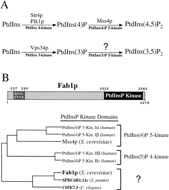
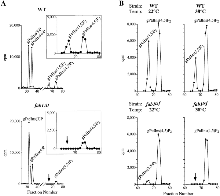
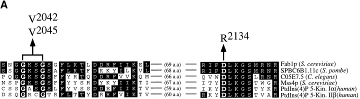
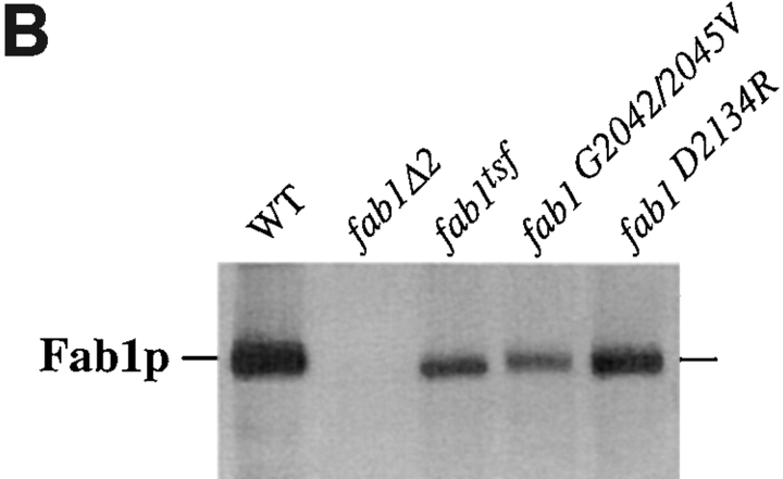
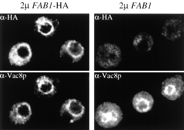
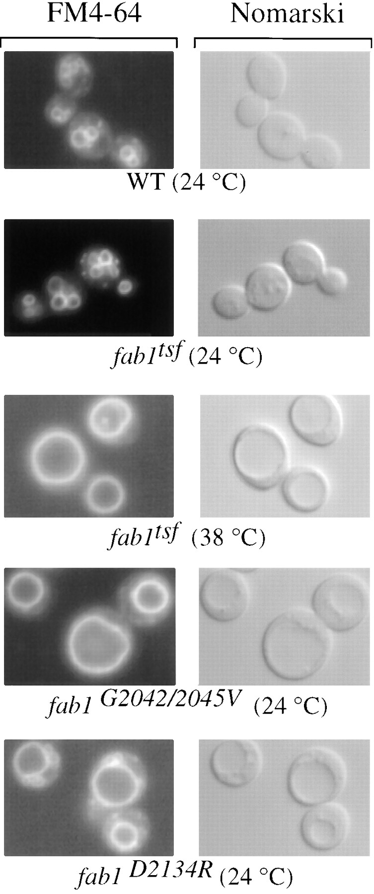
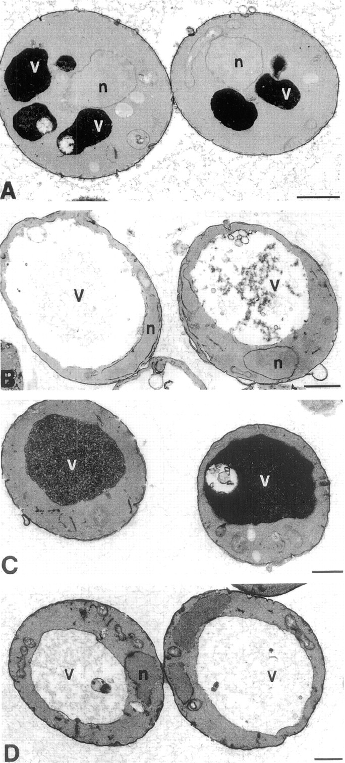
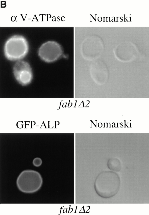
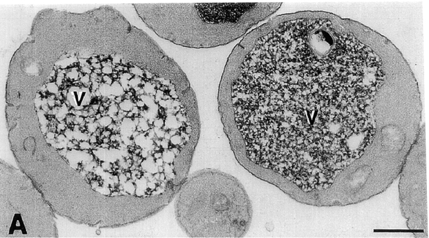
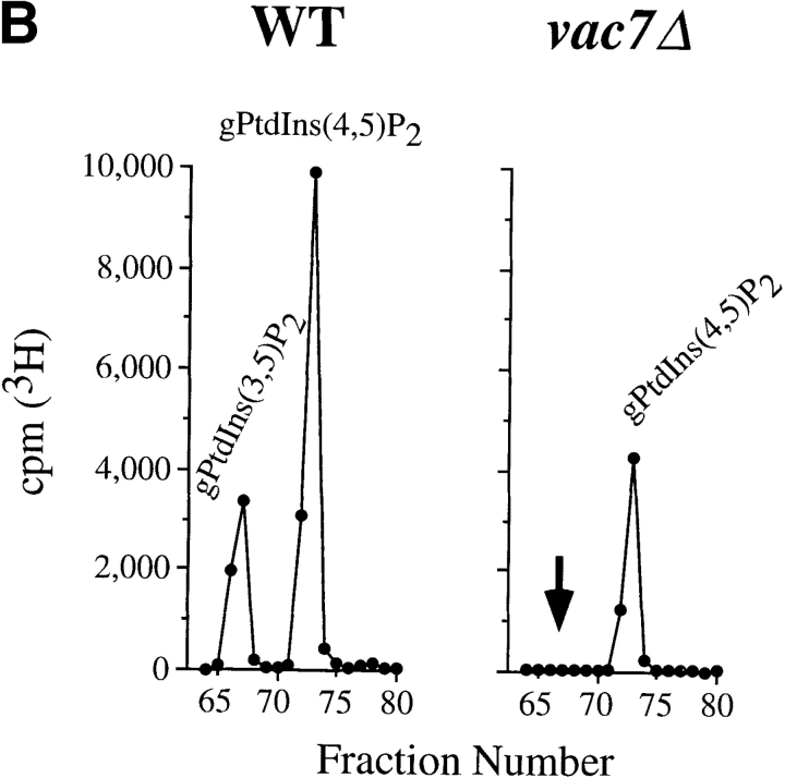
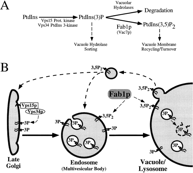
The Saccharomyces cerevisiae FAB1 gene encodes a 257-kD protein that contains a cysteine-rich RING-FYVE domain at its NH2-terminus and a kinase domain at its COOH terminus. Based on its sequence, Fab1p was initially proposed to function as a phosphatidylinositol 4-phosphate (PtdIns(4)P) 5-kinase (). Additional sequence analysis of the Fab1p kinase domain, reveals that Fab1p defines a subfamily of putative PtdInsP kinases that is distinct from the kinases that synthesize PtdIns(4,5)P2. Consistent with this, we find that unlike wild-type cells, fab1Delta, fab1(tsf), and fab1 kinase domain point mutants lack detectable levels of PtdIns(3,5)P2, a phosphoinositide recently identified both in yeast and mammalian cells. PtdIns(4,5)P2 synthesis, on the other hand, is only moderately affected even in fab1Delta mutants. The presence of PtdIns(3)P in fab1 mutants, combined with previous data, indicate that PtdIns(3,5)P2 synthesis is a two step process, requiring the production of PtdIns(3)P by the Vps34p PtdIns 3-kinase and the subsequent Fab1p- dependent phosphorylation of PtdIns(3)P yielding PtdIns(3,5)P2. Although Vps34p-mediated synthesis of PtdIns(3)P is required for the proper sorting of hydrolases from the Golgi to the vacuole, the production of PtdIns(3,5)P2 by Fab1p does not directly affect Golgi to vacuole trafficking, suggesting that PtdIns(3,5)P2 has a distinct function. The major phenotypes resulting from Fab1p kinase inactivation include temperature-sensitive growth, vacuolar acidification defects, and dramatic increases in vacuolar size. Based on our studies, we hypothesize that whereas Vps34p is essential for anterograde trafficking of membrane and protein cargoes to the vacuole, Fab1p may play an important compensatory role in the recycling/turnover of membranes deposited at the vacuole. Interestingly, deletion of VAC7 also results in an enlarged vacuole morphology and has no detectable PtdIns(3,5)P2, suggesting that Vac7p functions as an upstream regulator, perhaps in a complex with Fab1p. We propose that Fab1p and Vac7p are components of a signal transduction pathway which functions to regulate the efflux or turnover of vacuolar membranes through the regulated production of PtdIns(3,5)P2.


Figures

Figure 1
Figure 1
Phosphatidylinositol derivatives and their kinases in yeast. (A) A diagram of the D-3 and D-4 phosphorylated phosphoinositides present in the yeast S. cerevisiae. Above the arrows, the specific kinases determined to be involved in their biosynthesis are listed. (B) A phylogenetic comparison of the Fab1p lipid kinase domain to the analogous domain of other PtdInsP kinases. Using the DNAStar program MegAlign, the S. cerevisiae Fab1p kinase domain (a.a. 2022–2263) and its homologues SPBC6B1.11c (AL021838; a.a. 358–595) and C05E7.5 (Z67879; a.a. 924–1170) from S. pombe and C. elegans, respectively, were aligned and a phylogenetic tree was constructed with the corresponding kinase domains of human PtdIns(4)P 5-kinases Type Iα (U78575; a.a. 125–449), Type Iβ (U52376; a.a. 1–288), Type IIβ (U85245; a.a. 100–416), Type IIc (P53807; a.a. 110–406), and S. cerevisiae Mss4p (D13716; a.a. 439–765). The multiple sequence alignment was performed using the Jotun-Hein method with default program parameters, using the PAM250 residue weight table.
Figure 2
Figure 2
PtdIns(3,5)P2 is undetectable in fab1 mutant cells. (A) Wild-type (SEY6210) and fab1Δ1 cells were labeled with myo- [2-3H]inositol for 12 h at 22°C and hyperosmotically shocked with 0.9 M NaCl for 10 min. Cells were then glass bead lysed and the cellular lipids extracted, deacylated and separated by HPLC (see Materials and Methods). Fractions (0.67 min) were collected and counted for [3H] radioactivity. Deacylated (glycero-) PtdIns(3)P, PtdIns(4)P, PtdIns(3,5)P2, and PtdIns(4,5)P2 are indicated. Arrows indicate the expected position of gPtdIns(3,5)P2. (B) Wild-type and fab1tsf cells were labeled for 12 h with myo-[2-3H]inositol at 22°C, after which both cultures were separated into two equal aliquots. One aliquot of each strain was incubated with 0.9 M NaCl for 10 min at 22°C. The remaining wild-type and fab1tsf cells were incubated with 0.9 M NaCl for 5 min at 22°C and, for the final 5 min of hyperosmotic shock, shifted to the nonpermissive temperature of 38°C. The peaks representing PtdIns(3,5)P2 and PtdIns(4,5)P2 are indicated. The arrow highlights the elution point of the gPtdIns(3,5)P2 standard. The data presented are representative of multiple experiments.
Figure 3
Figure 3
Point mutations within the Fab1p kinase domain. (A) A conserved region of the Fab1p kinase domain is aligned with several lipid kinase homologues identified from a BLAST search. Residues identical to those in Fab1p are outlined in black. Above the alignment, the amino acid substitutions engineered to make the Fab1p point mutant constructs are shown. (B) Fab1p was immunoprecipitated from a wild-type (SEY6210), fab1Δ2, fab1tsf, as well as the two kinase domain point mutant strains (fab1G2042/2045V and fab1D2134R). Cells were metabolically labeled with Express [35S]-protein labeling mix and then chased in an excess of methionine and cysteine for 45 min at 24°C (see Materials and Methods). The immunoprecipitated protein was visualized after SDS-PAGE by fluorography (7-d exposure at −80°C).
Figure 3
Figure 3
Point mutations within the Fab1p kinase domain. (A) A conserved region of the Fab1p kinase domain is aligned with several lipid kinase homologues identified from a BLAST search. Residues identical to those in Fab1p are outlined in black. Above the alignment, the amino acid substitutions engineered to make the Fab1p point mutant constructs are shown. (B) Fab1p was immunoprecipitated from a wild-type (SEY6210), fab1Δ2, fab1tsf, as well as the two kinase domain point mutant strains (fab1G2042/2045V and fab1D2134R). Cells were metabolically labeled with Express [35S]-protein labeling mix and then chased in an excess of methionine and cysteine for 45 min at 24°C (see Materials and Methods). The immunoprecipitated protein was visualized after SDS-PAGE by fluorography (7-d exposure at −80°C).
Figure 4
Figure 4
fab1 point mutant strains are compromised for PtdIns(3,5)P2 synthesis. Wild-type (SEY6210), fab1G2042/2045V, and fab1D2134R cells were labeled with myo- [2-3H]inositol for 12 h at 24°C and hyperosmotically shocked with 0.9 M NaCl for 10 min (see Fig. 2). The deacylated PtdIns derivatives were separated by HPLC, only the region of the elution profile separating gPtdIns(3,5)P2 and gPtdIns(4,5)P2 are shown. The arrow highlights the elution point of the glycero-PtdIns(3,5)P2 standard.
Figure 5
Figure 5
Localization of HA-tagged Fab1p by indirect immunofluorescence. Cells overexpressing HA-tagged Fab1 or wild-type Fab1 protein from multi-copy plasmids were fixed, converted to spheroplasts and then incubated with monoclonal anti-HA or anti-Vac8p antibodies as described in the Materials and Methods. The localization of Fab1p-HA and Vac8p were determined by indirect immunofluorescence. HA-tagged Fab1p and Vac8p signals are indicated.
Figure 6
Figure 6
fab1 mutant cells have an abnormal vacuole morphology. In the right panels, vacuoles were observed by Nomarski optics. On the left, the same fields are shown under fluorescent illumination (rhodamine channel). Cells were grown to mid-log phase at 24°C and the vacuoles labeled with the vital stain FM4-64 for 15 min (see Materials and Methods). After labeling, all of the strains were chased in the absence of dye for 1 h at 24°C except a duplicate of the fab1tsf strain that was chased at the nonpermissive temperature of 38°C.
Figure 7
Figure 7
Electron microscopy of fab1 mutants. Cells were grown to mid-log phase and fixed with 3% glutaraldehyde. Cells were then converted to spheroplasts and visualized by electron microscopy (see Materials and Methods). (A) Wild-type (SEY6210) cells. (B) fab1Δ1 cells. (C) fab1G2042/2045V cells. (D) fab1D2134R cells. Bar, 0.5 μm.
Figure 8
Figure 8
Intracellular sorting of vacuolar hydrolases in fab1 mutant cells. Yeast cells were metabolically labeled with Express [35S]-protein labeling mix during a 10-min pulse and then chased in the presence of an excess of nonradiolabeled methionine and cysteine for 0, 15, or 30 min (see Materials and Methods). Each of the vacuolar enzymes was immunoprecipitated, followed by SDS-PAGE and autoradiography. The migration positions of the precursor and mature forms of the hydrolases are indicated on the left side of the figure panels. (A) The maturation of CPY and ALP was examined in the fab1tsf strain compared with wild-type, as described in Materials and Methods. However, for the nonpermissive temperature, cells were preshifted to 38°C for 30 min before labeling and maintained at the elevated temperature throughout the chase period. (B) The upper panel, on the left, shows indirect immunofluorescence localization of the 60-kD subunit of the vacuolar ATPase using a monoclonal antibody in the fab1Δ2 strain. For the immunofluorescence, cells were fixed overnight, spheroplasted, and then incubated with antibody as described in Materials and Methods. The lower panel, on the left, shows GFP-ALP localization in a fab1Δ2 strain, observed by fluorescence microscopy (see Materials and Methods). On the right side for both panels are the identical fields observed under Nomarski optics. (C) The maturation of the vacuolar hydrolases CPS and ALP in the two point mutant strains (fab1G2042/2045V and fab1D2134R) and the wild-type strain SEY6210. The labeling and chase were done as in A however, the cells were maintained at 24°C.
Figure 8
Figure 8
Intracellular sorting of vacuolar hydrolases in fab1 mutant cells. Yeast cells were metabolically labeled with Express [35S]-protein labeling mix during a 10-min pulse and then chased in the presence of an excess of nonradiolabeled methionine and cysteine for 0, 15, or 30 min (see Materials and Methods). Each of the vacuolar enzymes was immunoprecipitated, followed by SDS-PAGE and autoradiography. The migration positions of the precursor and mature forms of the hydrolases are indicated on the left side of the figure panels. (A) The maturation of CPY and ALP was examined in the fab1tsf strain compared with wild-type, as described in Materials and Methods. However, for the nonpermissive temperature, cells were preshifted to 38°C for 30 min before labeling and maintained at the elevated temperature throughout the chase period. (B) The upper panel, on the left, shows indirect immunofluorescence localization of the 60-kD subunit of the vacuolar ATPase using a monoclonal antibody in the fab1Δ2 strain. For the immunofluorescence, cells were fixed overnight, spheroplasted, and then incubated with antibody as described in Materials and Methods. The lower panel, on the left, shows GFP-ALP localization in a fab1Δ2 strain, observed by fluorescence microscopy (see Materials and Methods). On the right side for both panels are the identical fields observed under Nomarski optics. (C) The maturation of the vacuolar hydrolases CPS and ALP in the two point mutant strains (fab1G2042/2045V and fab1D2134R) and the wild-type strain SEY6210. The labeling and chase were done as in A however, the cells were maintained at 24°C.
Figure 9
Figure 9
The vac7Δ strain displays phenotypes similar to the fab1Δ1 strain. (A) Electron microscopy of vac7Δ cells prepared as in Fig. 6. (B) Wild-type and vac7Δ cells were labeled 12 h with myo-[2-3H]inositol and osmotically shocked with 0.9 M NaCl for 10 min at 22°C. Each strain was then lysed and the extracted cellular lipids deacylated and separated by HPLC as in Fig. 2. Deacylated products representing PtdIns(3,5)P2 and PtdIns(4,5)P2 are indicated. The arrow indicates the expected position of PtdIns(3,5)P2. The data are representative of several experiments. Bar, 0.5 μm.
Figure 9
Figure 9
The vac7Δ strain displays phenotypes similar to the fab1Δ1 strain. (A) Electron microscopy of vac7Δ cells prepared as in Fig. 6. (B) Wild-type and vac7Δ cells were labeled 12 h with myo-[2-3H]inositol and osmotically shocked with 0.9 M NaCl for 10 min at 22°C. Each strain was then lysed and the extracted cellular lipids deacylated and separated by HPLC as in Fig. 2. Deacylated products representing PtdIns(3,5)P2 and PtdIns(4,5)P2 are indicated. The arrow indicates the expected position of PtdIns(3,5)P2. The data are representative of several experiments. Bar, 0.5 μm.
Figure 10
Figure 10
(A) The pathway for the biosynthesis of PtdIns(3,5)P2 in yeast. The Vps15/34p-dependent phosphorylation of PtdIns leads to the production of PtdIns(3)P. A pool of this lipid then either traffics to the lumen of the vacuole for degradation or remains cytoplasmically exposed, serving as a substrate for a second, Fab1p kinase domain-dependent phosphotransfer reaction resulting in the synthesis of PtdIns(3,5)P2. Below the phosphorylated lipid products, roles for these lipids are indicated. PtdIns(3)P is required in anterograde traffic from the Golgi to the vacuole, whereas PtdIns(3,5)P2 may regulate the efflux of vacuolar membrane material. (B) A model for Fab1p-mediated regulation of vacuolar membrane recycling/turnover. Activation of Vps34p by the membrane associated lipid kinase Vps15p, leads to the site-specific synthesis of PtdIns(3)P at the Golgi/endosomal membrane. The production of PtdIns(3)P is not only required for this biosynthetic trafficking route (highlighted by the solid arrows), but also is used by the Fab1p lipid kinase for the synthesis of PtdIns(3,5)P2. A large pool of PtdIns(3)P is turned over in a hydrolase-dependent manner, possibly by being sorted into the inner leaflet of endosomal invaginations which are transported to the lumen of the vacuole and, together with PtdIns(3)P, degraded. A separate pool of PtdIns(3)P must remain in the cytoplasmic membrane leaflet in order to be available to Fab1p. The presence of PtdIns(3,5)P2 may then serve to recruit factors involved in the efflux of membrane to earlier compartments (top, dashed lines) or cause the internalization of vacuolar membrane into the lumen for degradation.

References

    1. Altschul SF, Madden TL, Schaffer AA, Zhang J, Zhang Z, Miller W, Lipman DJ. Gapped BLAST and PSI-BLAST: a new generation of protein database search programs. Nucleic Acids Res. 1997;25:3389–3402. - PMC - PubMed
    1. Auger KR, Carpenter CL, Cantley LC, Varticovski L. Phosphatidylinositol 3-kinase and its novel product, phosphatidylinositol 3-phosphate, are present in Saccharomyces cerevisiae. . J Biol Chem. 1989;264:20181–20184. - PubMed
    1. Babst M, Sato TK, Banta LM, Emr SD. Endosomal transport function in yeast requires a novel AAA-type ATPase, Vps4p. EMBO (Eur Mol Biol Organ) J. 1997;16:1820–1831. - PMC - PubMed
    1. Bankaitis VA, Aitken JR, Cleves AE, Dowhan W. An essential role for a phospholipid transfer protein in yeast Golgi function. Nature. 1990;347:561–562. - PubMed
    1. Banta LM, Robinson JS, Klionsky DJ, Emr SD. Organelle assembly in yeast: characterization of yeast mutants defective in vacuolar biogenesis and protein sorting. J Cell Biol. 1988;107:1369–1383. - PMC - PubMed

Publication types

MeSH terms

Substances