. 1998 Oct 5;143(1):217-24.
doi: 10.1083/jcb.143.1.217.

Bax-induced cytochrome C release from mitochondria is independent of the permeability transition pore but highly dependent on Mg2+ ions

Affiliations

Bax-induced cytochrome C release from mitochondria is independent of the permeability transition pore but highly dependent on Mg2+ ions

R Eskes et al. J Cell Biol. .

Abstract

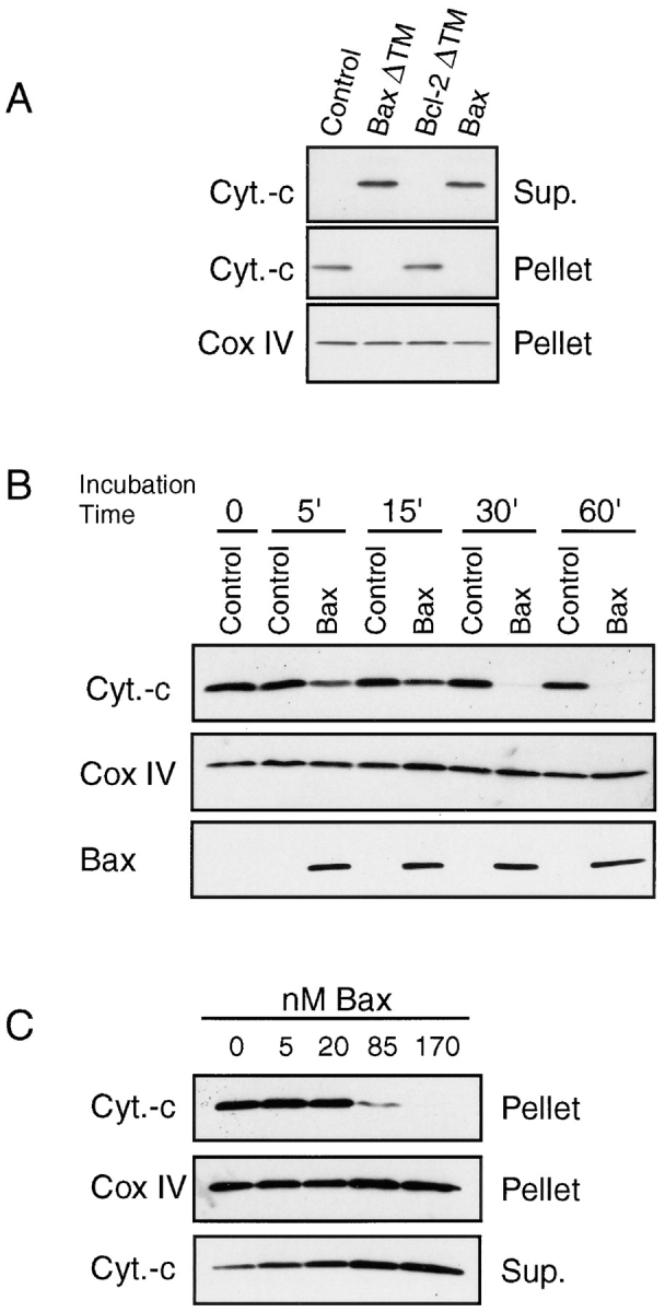
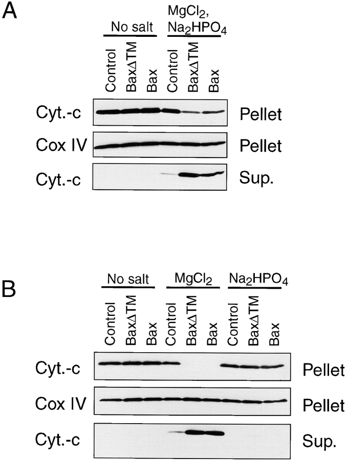
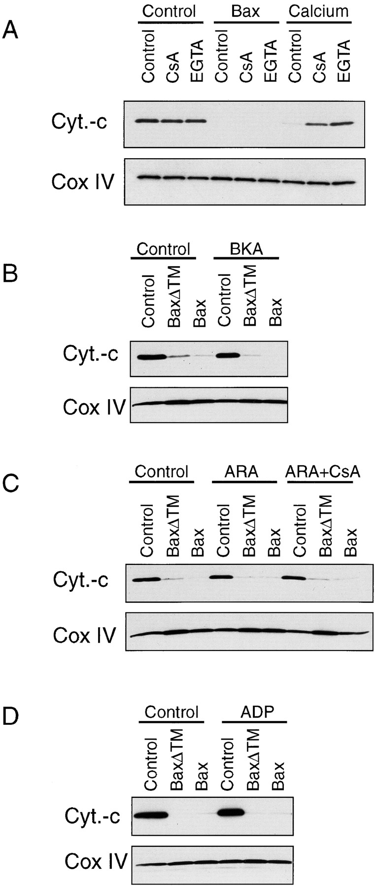
Bcl-2 family members either promote or repress programmed cell death. Bax, a death-promoting member, is a pore-forming, mitochondria-associated protein whose mechanism of action is still unknown. During apoptosis, cytochrome C is released from the mitochondria into the cytosol where it binds to APAF-1, a mammalian homologue of Ced-4, and participates in the activation of caspases. The release of cytochrome C has been postulated to be a consequence of the opening of the mitochondrial permeability transition pore (PTP). We now report that Bax is sufficient to trigger the release of cytochrome C from isolated mitochondria. This pathway is distinct from the previously described calcium-inducible, cyclosporin A-sensitive PTP. Rather, the cytochrome C release induced by Bax is facilitated by Mg2+ and cannot be blocked by PTP inhibitors. These results strongly suggest the existence of two distinct mechanisms leading to cytochrome C release: one stimulated by calcium and inhibited by cyclosporin A, the other Bax dependent, Mg2+ sensitive but cyclosporin insensitive.
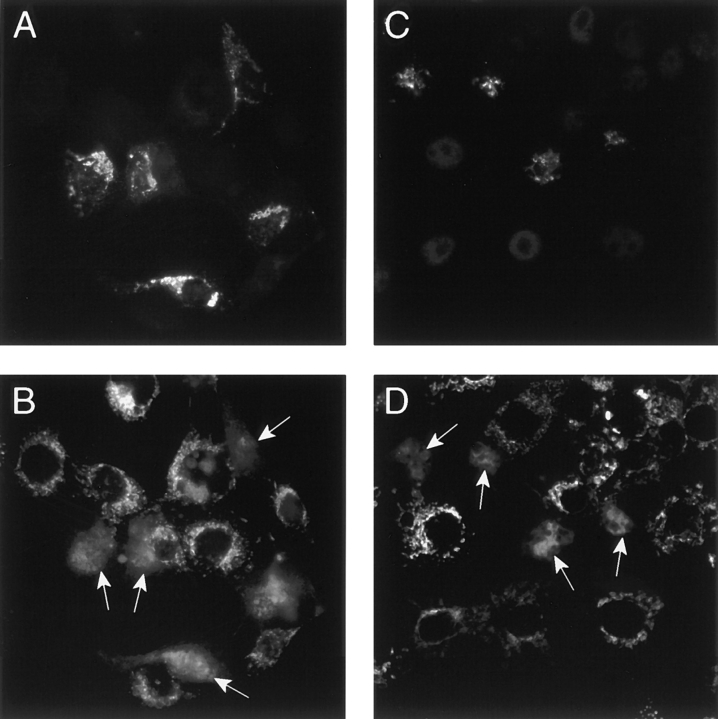

Figures

Figure 1
Figure 1
Mitochondrial localization of Bax. HeLa cells were transiently transformed with cDNA encoding a His-tagged Bax and immunostained 15 h later for both the mitochondrial marker HSP-70 (A) and Bax (B). Note that the shape as well as the distribution of the mitochondria has change in the Bax-transfected cell (arrow).
Figure 2
Figure 2
Immunofluorescence studies of cytochrome C release from mitochondria after overexpression of Bax in HeLa cells. HeLa cells were transiently transfected with a cDNA encoding a His-tagged human Bax and cultured for 15 h in the presence (D–F) or absence (A–C) of z-VAD-fmk before immunostaining for Bax (A and D) and cytochrome C (B and E). C and F are nuclear Hoechst stainings. Arrows, transfected cells.
Figure 3
Figure 3
Bax-induced cytochrome C release from isolated mitochondria. (A) Mitochondria isolated from mouse liver were incubated for 1 h at 30°C with 5 μM COOH-terminal truncated Bax (BaxΔTM), 5 μM COOH-terminal truncated Bcl-2 or 170 nM full-length Bax in 200 μl KCl buffer. After 1 h, the reaction mixture was centrifuged at 13,000 g for 5 min. Supernatant and mitochondrial pellets corresponding to 5 μg mitochondrial proteins were subjected to 4–20% SDS-PAGE and analyzed by Western blot. Loading for the mitochondrial pellet was controlled with a Cox IV antibody. (B) Kinetics of Bax-induced cytochrome C release. Mitochondria were incubated with 170 nM Bax or buffer control at 30°C and the mitochondria pellet was analysed over time as in A. In addition to cytochrome C and Cox IV, Bax levels were measured in the mitochondrial pellets. (C) Mitochondria were incubated with different amounts of Bax for 1 h at 30°C and cytochrome C release was measured as in A. Data shown are representative of 3–12 independent experiments performed with at least three different preparations of Bax protein.
Figure 4
Figure 4
Bax effect is ion dependent. Mitochondria isolated from mouse liver cells were incubated with 170 nM Bax at 30°C in sucrose-mannose buffer with or without addition of salts as indicated in the figure. The salt concentrations were 4 mM MgCl2 and 5 mM Na2HPO4. Cytochrome C release was measured after 1 h as described in Fig. 3.
Figure 5
Figure 5
Bax-induced release of cytochrome C from isolated mitochondria is not inhibited by PTP blockers. Several PTP blockers have been tested for their ability to inhibit the release of cytochrome C from isolated mitochondria induced by 170 nM Bax. (A) We compared Bax effect with that of calcium, a PTP opener, and their sensitivity to 10 μM CsA and 0.5 mM EGTA. Cytochrome C and Cox IV in the mitochondrial pellet were measured after a 1-h incubation in KCl buffer at 30°C by Western blotting. The PTP inhibitors BKA (100 μM) (B), a combination of CsA (10 μM) and ARA (50 μM) (C) and ADP (10 μM) (D) were also tested. Mitochondria were preincubated with these compounds for 5 min before addition of Bax. Note that although CsA and EGTA were capable of inhibiting the calcium effect, they failed to block Bax-induced cytochrome C release.
Figure 6
Figure 6
Both Cyclosporin A and BKA fail to inhibit Bax-induced release of cytochrome C in COS cells. COS cells were transfected with a cDNA encoding His-Bax and cultured for 15 h in the presence of 10 μM CsA and 100 μM z-VAD-fmk (A and B) or 100 μM BKA and 100 μM z-VAD-fmk (C and D) before immunostaining for Bax (A and C) and cytochrome C (B and D). Note that all cells that overexpress Bax display a diffuse cytosolic cytochrome C immunostaining. Arrows, transfected cells.

References

    1. Antonson B, Conti F, Ciavatta AM, Montesuit S, Lewis S, Martinou I, Bernasconi L, Bernard A, Mermod J-J, Mazzei G, Maundrell K, Gambale F, Sadoul R, Martinou J-C. Inhibition of Bax channel forming activity by Bcl-2. Science. 1997;277:370–372. - PubMed
    1. Bernardi P, Petronilli V. The permeability transition pore as a mitochondrial calcium release channel; a critical appraisal. J Bioenerg Biomembr. 1996;28:129–136. - PubMed
    1. Beutner G, Rueck A, Riede B, Welte W, Brdiczka D. Complexes between kinases, mitochondrial porin, and adenylate tranlocator in rat brain resemble the permeability transition pore. FEBS (Fed Eur Biochem Soc) Lett. 1996;396:189–195. - PubMed
    1. Bossy-Wetzel E, Newmeyer DD, Green DR. Mitochondrial cytochrome C release in apoptosis occurs upstream of DEVD-specific caspase activation and independently of mitochondrial transmembrane depolarization. EMBO (Eur Mol Biol Organ) J. 1998;17:37–49. - PMC - PubMed
    1. Boyer PD. The ATP synthase—a splendid molecular machine. Annu Rev Biochem. 1997;66:717–749. - PubMed

MeSH terms