. 1998 Oct;9(10):2905-16.
doi: 10.1091/mbc.9.10.2905.

Activation and cellular localization of the cyclosporine A-sensitive transcription factor NF-AT in skeletal muscle cells

Affiliations
Free PMC article

Activation and cellular localization of the cyclosporine A-sensitive transcription factor NF-AT in skeletal muscle cells

K L Abbott et al. Mol Biol Cell. 1998 Oct.
Free PMC article

Abstract

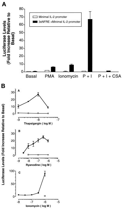
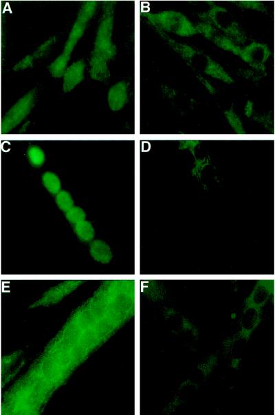
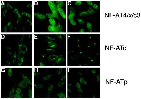
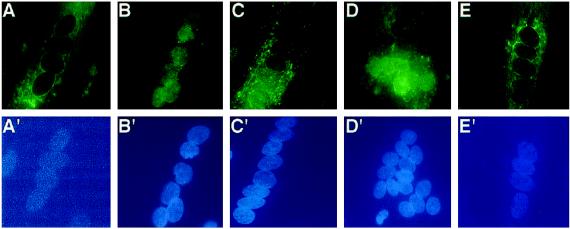
The widely used immunosuppressant cyclosporine A (CSA) blocks nuclear translocation of the transcription factor, NF-AT (nuclear factor of activated T cells), preventing its activity. mRNA for several NF-AT isoforms has been shown to exist in cells outside of the immune system, suggesting a possible mechanism for side effects associated with CSA treatment. In this study, we demonstrate that CSA inhibits biochemical and morphological differentiation of skeletal muscle cells while having a minimal effect on proliferation. Furthermore, in vivo treatment with CSA inhibits muscle regeneration after induced trauma in mice. These results suggest a role for NF-AT-mediated transcription outside of the immune system. In subsequent experiments, we examined the activation and cellular localization of NF-AT in skeletal muscle cells in vitro. Known pharmacological inducers of NF-AT in lymphoid cells also stimulate transcription from an NF-AT-responsive reporter gene in muscle cells. Three isoforms of NF-AT (NF-ATp, c, and 4/x/c3) are present in the cytoplasm of muscle cells at all stages of myogenesis tested. However, each isoform undergoes calcium-induced nuclear translocation from the cytoplasm at specific stages of muscle differentiation, suggesting specificity among NF-AT isoforms in gene regulation. Strikingly, one isoform (NF-ATc) can preferentially translocate to a subset of nuclei within a single multinucleated myotube. These results demonstrate that skeletal muscle cells express functionally active NF-AT proteins and that the nuclear translocation of individual NF-AT isoforms, which is essential for the ability to coordinate gene expression, is influenced markedly by the differentiation state of the muscle cell.


Figures

Figure 1
Figure 1
CSA inhibits myoblast differentiation with little effect on proliferation. (A) CSA minimally inhibits DNA synthesis in myoblasts. Human myoblasts were treated with different doses of CSA for 24–72 h. The cells were pulse labeled with 3H-thymidine for 2 h, and the number of TCA-precipitable counts per min was determined. The data are plotted as the amount of 3H-thymidine incorporation in the drug-treated cells as a percent of that in vehicle-treated cells. No difference was noted among the different treatment times, so the data for all timepoints were pooled. Each point represents the mean ± SD of four experiments each performed in triplicate. (B) CSA inhibits expression of embryonic myosin heavy chain, a marker of differentiated muscle cells, in a dose-dependent manner. Top panel, immunoblots of electrophorectically separated proteins of vehicle and CSA- treated human muscle cells for 48–80 h in fusion medium using an antibody to EMyHC are shown. Detection was with enhanced luminescence. Representative blot of three experiments is shown. Bottom panel, immunoblots were scanned and quantitated using NIH Image. Each point represents the mean ± SEM of EMyHC expression in CSA-treated samples relative to vehicle in three experiments, each performed in duplicate. (C) CSA inhibits CK enzyme activity in a dose-dependent manner. CK activity was determined in samples treated for 24 h in ITS FM. Each point represents the mean ± SEM of differentiation-specific CK activity in CSA-treated samples relative to vehicle in three experiments, each performed in triplicate. (D) CSA inhibits myoblast fusion both in vitro and in vivo. Top panels, high-density mouse myoblasts were treated either with vehicle (panel A) or 10−6 M CSA (panel B) for 24 h in ITS fusion medium. Few multinucleated myotubes are present in CSA-treated cultures. Bar, 20 μm. Bottom panels, CSA inhibits muscle regeneration in vivo. Localized damage was induced in the tibialis anterior muscles of mice. Groups of mice were either treated with vehicle (panel C) or 45 mg/kg CSA i.p. (panel D) daily for 10 d. Muscle from vehicle-treated animals is characterized by centrally nucleated muscle fibers of various sizes as expected at this timepoint. Centrally nucleated muscle fibers are a hallmark of muscle regeneration. Muscle from CSA-treated animals is greatly deficient in regenerated muscle fibers. Cryostat sections of muscle in cross-section stained with hematoxylin and eosin are shown.
Figure 2
Figure 2
Induction of NF-AT–mediated transcription in skeletal muscle cells. (A) Myotubes contain NF-AT proteins that are functionally active in response to known pharmacological inducers of NF-AT–mediated transcription in T cells. Primary mouse myotubes containing either the control (minimal IL-2 promoter only, □) or the NF-AT-responsive (triplex of the distal IL-2 gene NF-AT response element [NFRE] upstream of the minimal IL-2 promoter,▪) retroviral reporter plasmids were treated for 6 h with vehicle alone (basal), PMA (10 nM), or ionomycin (1 μM) alone or the two together (P+I). Some cultures were pretreated with cyclosporine A (CSA; 1 μM) for 1 h before addition of PMA and ionomycin to the medium. Luciferase values were subsequently measured. Luciferase activity is induced only in the cells containing the NF-AT response element upstream of the minimal IL-2 promoter. Both PMA and ionomycin stimulate NF-AT–mediated transcription in myotubes, but the two drugs together markedly syngerize. The P+I response is blocked in the presence of CSA. Each bar represents the mean ± SEM of two experiments, each performed in triplicate. (B) Drugs with different mechanisms of increasing intracellular calcium induce NF-AT– mediated transcription in myotubes. Primary mouse myotubes containing the NF-AT responsive reporter plasmid were treated with vehicle alone or different doses of thapsigargin (panel A), ryanodine (panel B), or ionomycin (panel C) in the presence of 10 nM PMA for 6 h before the measurement of luciferase activity. Cultures were either treated with these drugs alone (•) or together with 1 μM CSA (○). All three agents induce NF-AT–mediated transcription, but the maximum levels of induction differ, with ionomycin giving the greatest induction. Each point represents the mean ± SEM of three independent experiments, each performed in triplicate.
Figure 3
Figure 3
Induction of NF-AT–mediated transcription in muscle cells is developmentally regulated. (A) Primary mouse muscle cells were treated for 6 h with either vehicle alone or PMA (10 nM) and ionomycin (1 μM) together before the measurement of luciferase activity at the following stages of muscle development: myoblasts (Mb), nascent myotubes (N Mt), mature myotubes (M Mt). A twofold induction is observed in myoblasts, with the values progressively increasing to 10-fold and 19-fold in nascent and mature myotubes, respectively, as differentiation increases. Note that the observed level of induction in mature myotubes with PMA and ionomycin treatment varies among batches of retrovirally infected muscle cells (compare with 70-fold induction in Figure 2). Each bar represents the mean ± SEM of three independent experiments each performed in triplicate. (B) Muscle differentiation is not associated with an increase in the level of NF-AT proteins. Immunoblots were performed on electrophoretically separated proteins from myoblasts (Mb), nascent myotubes (NMt), and mature myotubes (MMt) using isoform-specific NF-AT antibodies to show that the increased levels of induction obtained with muscle differentiation are due to developmental differences and not simply protein levels.
Figure 4
Figure 4
Specificity of antibodies in immunohistochemistry of skeletal muscle cells. Human myoblasts (A and B) or myotubes (C and D) were treated with 10 nM thapsigargin for 1 h to concentrate the NF-AT proteins in the nucleus. Cells were fixed and stained with antibodies for NF-AT4/x/c3 (A and B) or NF-ATc (C and D) that had been preabsorbed with control extracts (A and C) or extracts from HEK cells transfected with specific NF-AT expression plasmids (B and D). For each antibody, the nuclear staining is eliminated by the extracts containing the specific NF-AT protein (B and D). The specificity of the NF-ATp antibody is shown by intense cytoplasmic staining of wild-type mouse muscle cells (E) but not of muscle cells from NF-ATp null mice (F).
Figure 5
Figure 5
Only one NF-AT isoform undergoes calcium-dependent nuclear translocation in mononucleated myoblasts. Human myoblasts were analyzed at near confluence for the expression of NF-AT4/x/c3, NF-ATc, or NF-ATp using isoform-specific antibodies and immunohistochemistry. Cells are shown with vehicle (A, D, and G), 10 nM thapsigargin for 1 h (B, E, and H), or 1 μM CSA pretreatment before thapsigargin treatment (C, F, and I). The nuclear translocation of NF-AT4/x/c3 (top panels) observed with thapsigargin treatment (B) is blocked in the presence of CSA (C). Nuclear translocation of NF-AT 4/x/c3 is detected in 90–95% of the myoblasts. Under similar conditions, NF-ATc (middle panels) and NF-ATp (bottom panels) do not undergo nuclear translocation. The data shown are representative of three experiments.
Figure 6
Figure 6
Calcium-dependent nuclear translocation of NF-ATp occurs only in nascent multinucleated myotubes. Human myotubes formed after 24 h in FM were treated with vehicle (panels A and A′), 10 nM thapsigargin (panels B and B′), or 10 nM thapsigargin and 1 μM CSA (panels C and C′) for 1 h before fixation and reaction with an antibody against NF-ATp. Top panels, immunofluorescent detection of NF-ATp in multinucleated myotubes; bottom panels, location of the individual nuclei in the same cell using the fluorescent DNA due, H33258. In nascent myotubes treated with vehicle (A), NF-ATp is cytoplasmic, but undergoes calcium-dependent nuclear translocation in 50–60% of myotubes (B), which is blocked by CSA (C). No nuclear selectivity is observed among the nuclei present within individual myotubes for NF-ATp. In mature myotubes (75–80 h in FM), NF-ATp does not undergo nuclear translocation in response to thapsigargin. The data shown are representative of two experiments.
Figure 7
Figure 7
NF-ATc can preferentially translocate to a subset of nuclei in multinucleated myotubes in response to calcium. NF-ATc undergoes calcium-dependent nuclear translocation in 90–95% of both nascent (24 h in FM) and mature (75–80 h in FM) human myotubes. Top panels, immunofluorescent detection of NF-ATc in multinucleated myotubes; bottom panels, location of the individual nuclei in the same cell using the fluorescent DNA due, H33258. In myotubes at both stages, NF-ATc is cytoplasmic (A). Thapsigargin treatment results in two types of nuclear translocation within the same population of cells. In some cells, NF-ATc translocates to all the nuclei within a single multinucleated myotube (B). In other cells, nuclear translocation is specific for a subset of nuclei within a single multinucleated myotube (C and D). CSA treatment blocks the nuclear translocation of NF-ATc in all myotubes (E). The data shown are representative of three experiments.

References

    1. Berman SA, Bursztajn S, Bowen B, Gilbert W. Localization of an acetylcholine receptor intron to the nuclear membrane. Science. 1990;247:212–214. - PubMed
    1. Boss V, Abbott KA, Wang X-F, Pavlath GK, Murphy TJ. Expression of the cyclosporin A-sensitive factor NFAT in cultured vascular smooth muscle cells: differential induction of NFAT-mediated transcription by phospholipase C-coupled cell surface receptors. J Biol Chem. 1998;273:19664–19671. - PubMed
    1. Boss V, Talpade DJ, Murphy TJ. Induction of NFAT-mediated transcription by Gq-coupled receptors in lymphoid and non-lymphoid cells. J Biol Chem. 1996;271:10429–10432. - PubMed
    1. Bradford MM. A rapid and sensitive method for the quantitation of microgram quantities of protein utilizing the principle of protein-dye binding. Anal Biochem. 1976;72:248–254. - PubMed
    1. Changeux JP. Compartmentalized transcription of acetylcholine receptor genes during motor endplate epigenesis. New Biol. 1991;3:413–429. - PubMed

Publication types

MeSH terms