

Electrochemical analysis of protein nitrotyrosine and dityrosine in the Alzheimer brain indicates region-specific accumulation
- PMID: 9763459
- PMCID: PMC6792852
- DOI: 10.1523/JNEUROSCI.18-20-08126.1998
Electrochemical analysis of protein nitrotyrosine and dityrosine in the Alzheimer brain indicates region-specific accumulation
Abstract
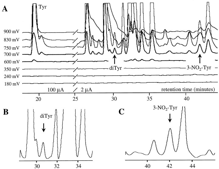
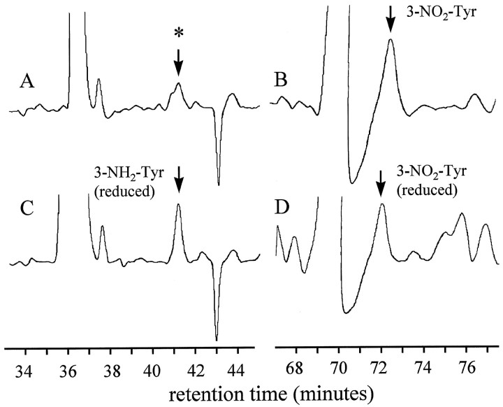
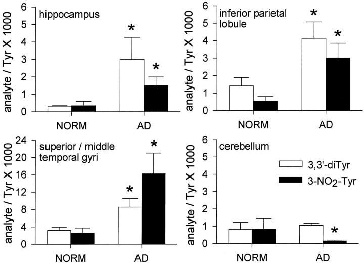
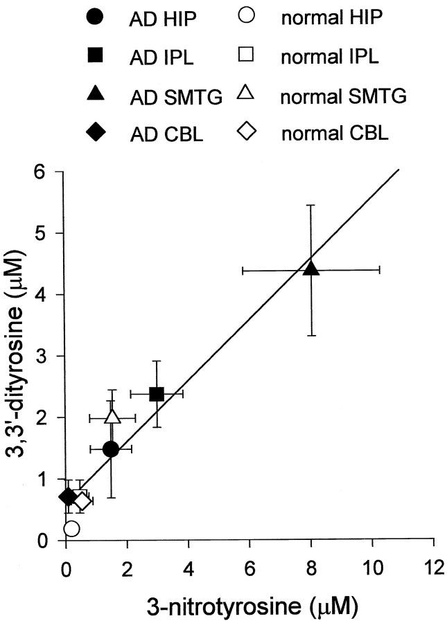
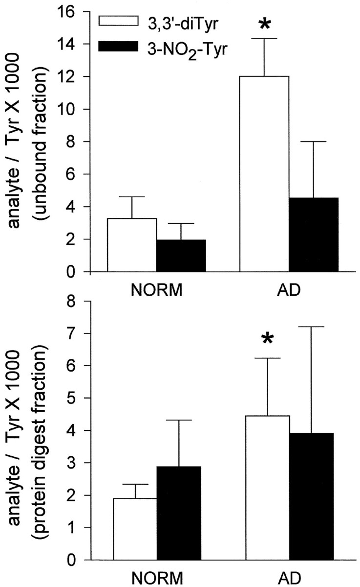
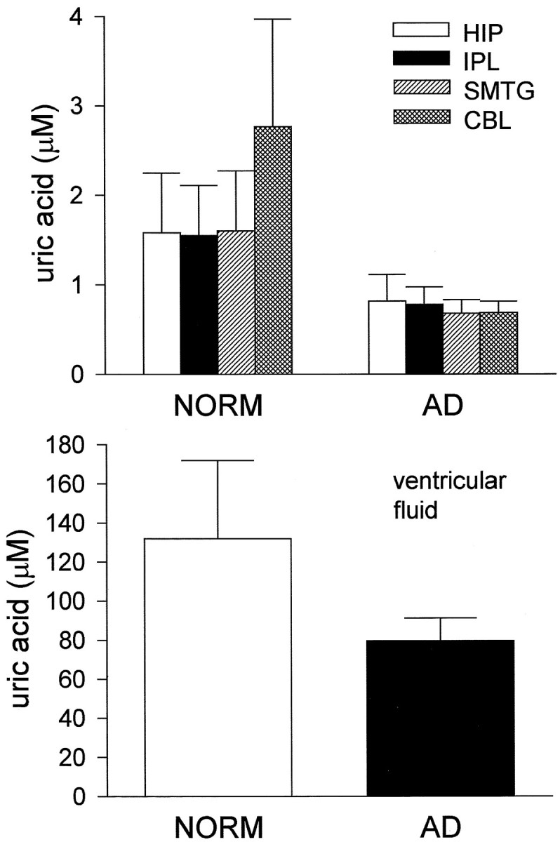
HPLC with electrochemical array detection (HPLC-ECD) was used to quantify 3,3'-dityrosine (diTyr) and 3-nitrotyrosine (3-NO2-Tyr) in four regions of the human brain that are differentially affected in Alzheimer's disease (AD). DiTyr and 3-NO2-Tyr levels were elevated consistently in the hippocampus and neocortical regions of the AD brain and in ventricular cerebrospinal fluid (VF), reaching quantities five- to eightfold greater than mean concentrations in brain and VF of cognitively normal subjects. Uric acid, a proposed peroxynitrite scavenger, was decreased globally in the AD brain and VF. The results suggest that AD pathogenesis may involve the activation of oxidant-producing inflammatory enzyme systems, including nitric oxide synthase.
Figures
References
-
- Ahlskog JE, Uitti RJ, Low PA, Tyce GM, Nickander KK, Peterson RC, Kokmen E. No evidence for systemic oxidant stress in Parkinson’s or Alzheimer’s disease. Mov Disord. 1995;10:566–573. - PubMed
-
- Balazs L, Leon M. Evidence of an oxidative challenge in the Alzheimer’s brain. Neurochem Res. 1994;19:1131–1137. - PubMed
-
- Bauer J, Strauss S, Schreiter-Gasser U, Ganter U, Schlegel P, Witt I, Volk B, Berger M. Interleukin 6 and α-2-macroglobulin indicate an acute-phase state in Alzheimer’s disease cortices. FEBS Lett. 1991;285:111–114. - PubMed
-
- Beckman JS, Chen J, Ischiropoulos H, Crow JP. Oxidative chemistry of peroxynitrite. Methods Enzymol. 1994;233:229–240. - PubMed
-
- Braak H, Braak E. Pathology of Alzheimer’s disease. In: Calne DB, editor. Neurodegenerative diseases. Saunders; Philadelphia: 1994. pp. 585–613.
Publication types
MeSH terms
Substances
Grants and funding
LinkOut - more resources
Full Text Sources
Other Literature Sources
Medical