. 1998 Oct 15;18(20):8300-10.
doi: 10.1523/JNEUROSCI.18-20-08300.1998.

Three-dimensional structure and composition of CA3-->CA1 axons in rat hippocampal slices: implications for presynaptic connectivity and compartmentalization

Affiliations

Three-dimensional structure and composition of CA3-->CA1 axons in rat hippocampal slices: implications for presynaptic connectivity and compartmentalization

G M Shepherd et al. J Neurosci. .

Abstract

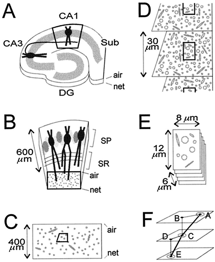
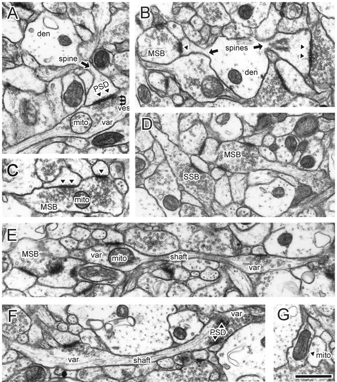
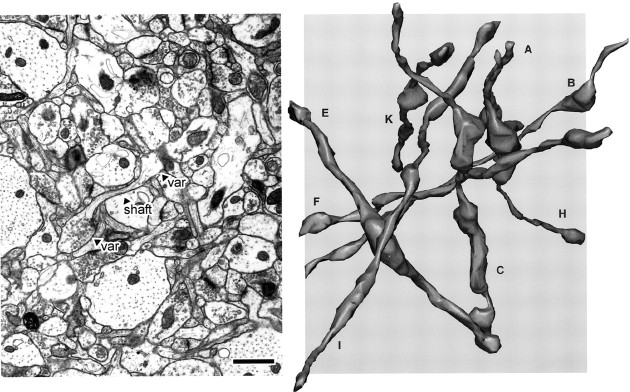
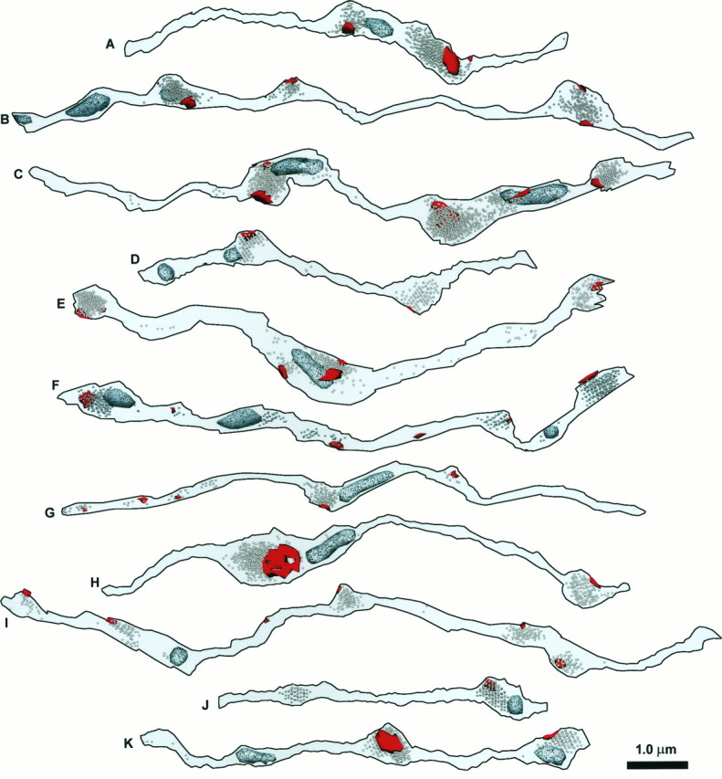
Physiological studies of CA3-->CA1 synaptic transmission and plasticity have revealed both pre- and postsynaptic effects. Understanding the extent to which individual presynaptic axonal boutons could provide local compartments for control of synaptic efficacy and microconnectivity requires knowledge of their three-dimensional morphology and composition. In hippocampal slices, serial electron microscopy was used to examine a nearly homogeneous population of CA3-->CA1 axons in the middle of stratum radiatum of area CA1. The locations of postsynaptic densities (PSDs), vesicles, and mitochondria were determined along 75 axon segments (9.1 +/- 2.0 micrometer in length). Synapses, defined by the colocalization of PSDs and vesicles, occurred on average at 2.7 micrometer intervals along the axons. Most varicosities (68%) had one PSD, 19% had 2-4 PSDs, and 13% had none. Synaptic vesicles occurred in 90% of the varicosities. One-half (53%) of the varicosities lacked mitochondria, raising questions about their regulation of ATP and Ca2+, and 8% of varicosities contained only mitochondria. Eleven axons were reconstructed fully. The varicosities were oblong and varied greatly in both length (1.1 +/- 0.7 micrometer) and volume (0.13 +/- 0.14 micrometer 3), whereas the intervaricosity shafts were narrow, tubular, and similar in diameter (0.17 +/- 0.04 micrometer) but variable in length (1.4 +/- 1.2 micrometer). The narrow axonal shafts resemble dendritic spine necks and thus could promote biochemical compartmentalization of individual axonal varicosities. The findings raise the intriguing possibility of localized differences in metabolism and connectivity among different axons, varicosities, and synapses.


Figures

Fig. 1.
Fig. 1.
Location, orientation, and sectioning of CA1 stratum radiatum samples. The air and netsurfaces are labeled. A, A hippocampal slice, depicting the Schaffer collateral of a CA3 pyramidal neuron projecting to the apical dendrites of a pyramidal neuron in CA1 stratum radiatum. After fixation, the slice was trimmed initially to leave just the central part of area CA1 (bold outline). DG, Dentate gyrus; Sub, subiculum. B, Higher power view of the trimmed area of CA1, showing pyramidal neurons in stratum pyramidale (SP) and their apical dendrites, which are cross-sectioned in the middle third of stratum radiatum (bold outline). C, Thick and thin sections of the block face, spanning the entire thickness of the slice, were examined to ascertain excellent tissue preservation and to determine the optimal level for taking serial thin sections. Then a right-angled trapezoid (bold outline) for serial thin sectioning was created at that level by trimming away surrounding tissue. D, On each serial thin section, micrographs were taken of the same region (bold outline).E, The resulting stack of serial micrographs totaled 107–109 images, with the dimensions indicated. F, The triangulation method used to measure lengths of axons. Thepoints at which the axons exited the stack of images, labeled A and E, and crossed the middle, labeled C, were marked. On overlays, the distances in the x–y plane between the exit points and the middle point were measured (line segments AB andCD). The z-axis differences between the points (line segments BC and DE) were calculated from the measured average section thickness and the number of intervening sections. The sum of the calculated hypotenuses (line segments AC and CE) gave the total length of the axon segment.
Fig. 2.
Fig. 2.
Ultrastructural features of CA3 varicosities and axons in stratum radiatum of area CA1. A, Synaptic bouton synapsing with a dendritic spine. B, A dendrite with two spines receiving input from two boutons, including one MSB. The MSB contained a mitochondrion in nearby sections (data not shown).C, Example of an MSB, with a presynaptic mitochondrion. Inspection of adjacent sections revealed a third PSD. D, Several boutons, including an MSB and SSB. The MSB, but not the SSB, contained out-of-plane mitochondria. E, Longitudinally sectioned axon with two boutons, neither of which had additional PSDs or mitochondria in adjacent images. F, Longitudinally sectioned axon. A mitochondrion occupied the right, but not the left, bouton when it was examined three-dimensionally.G, A varicosity occupied by a single mitochondrion but not associated with PSDs or vesicles. Note that the images shown in the subsequent figures are from different axons. den, Dendritic shaft; mito, mitochondria; MSB, multiple-synapse bouton; PSD, postsynaptic density,filled triangles in B andC; SSB, single-synapse bouton;shaft, axonal shaft (arrows);var, axonal varicosity; ves, vesicles. Scale bar, 1.0 μm.
Fig. 3.
Fig. 3.
Left, Full-field view of a representative micrograph from the stack of images used for three-dimensional reconstructions. The shaft and two varicosities (var) of an axon are indicated. Right, Eight reconstructed axons from series LMTN, at the same scale as the micrograph. Some axons extend beyond the area of the micrograph because of the three-dimensional perspective. The axons travel in many different directions, rather than parallel to each other. Scale bar, 1.0 μm.
Fig. 4.
Fig. 4.
The membrane contours for 11 reconstructed axon segments, labeled A–K, including eight from series LMTN and three from ZNQB. Axon segments were rotated from their various native positions, as seen in Figure 3, into uniform orientation.
Fig. 5.
Fig. 5.
The PSDs (red), vesicles, and mitochondria (speckled, light blue) for the same set of 11 reconstructed axon segments that are shown in Figure4. When they are photocopied, PSDs appear relatively smooth and black, whereas mitochondria are speckled and lighter.
Fig. 6.
Fig. 6.
Graph of measured lengths versus volumes for axonal shafts (filled circles) and varicosities (open circles). The dotted linerepresents the theoretical length–volume relationship for spherical varicosities; i.e., volume = (4/3) πr3, where r = (varicosity length)/2. Both shafts and varicosities are essentially tubular.

References

    1. Alnaes E, Rahamimoff R. On the role of mitochondria in transmitter release from motor nerve terminals. J Physiol (Lond) 1975;248:285–306. - PMC - PubMed
    1. Andersen P. Organization of hippocampal neurons and their interconnections. In: Isaacson RL, Pribram KH, editors. The hippocampus, Vol I. Plenum; New York: 1975. pp. 155–175.
    1. Andersen P, Bliss TVP, Skrede KK. Lamellar organization of hippocampal excitatory pathways. Exp Brain Res. 1971;13:222–238. - PubMed
    1. Andersen P, Trommald M, Jensen V. Low synaptic convergence of CA3 collaterals on CA1 pyramidal cells suggests few release sites. In: Stjärne L, Greengard P, Grillner S, Hökfelt T, Ottoson D, editors. Molecular and cellular mechanisms of neurotransmitter release. Raven; New York: 1994. pp. 341–351. - PubMed
    1. Fawcett DW. The cell. Saunders; Philadelphia: 1981.

Publication types

MeSH terms

LinkOut - more resources