. 1998 Oct 15;18(20):8485-95.
doi: 10.1523/JNEUROSCI.18-20-08485.1998.

Nicotine selectively enhances NMDA receptor-mediated synaptic transmission during postnatal development in sensory neocortex

Affiliations

Nicotine selectively enhances NMDA receptor-mediated synaptic transmission during postnatal development in sensory neocortex

V B Aramakis et al. J Neurosci. .

Abstract

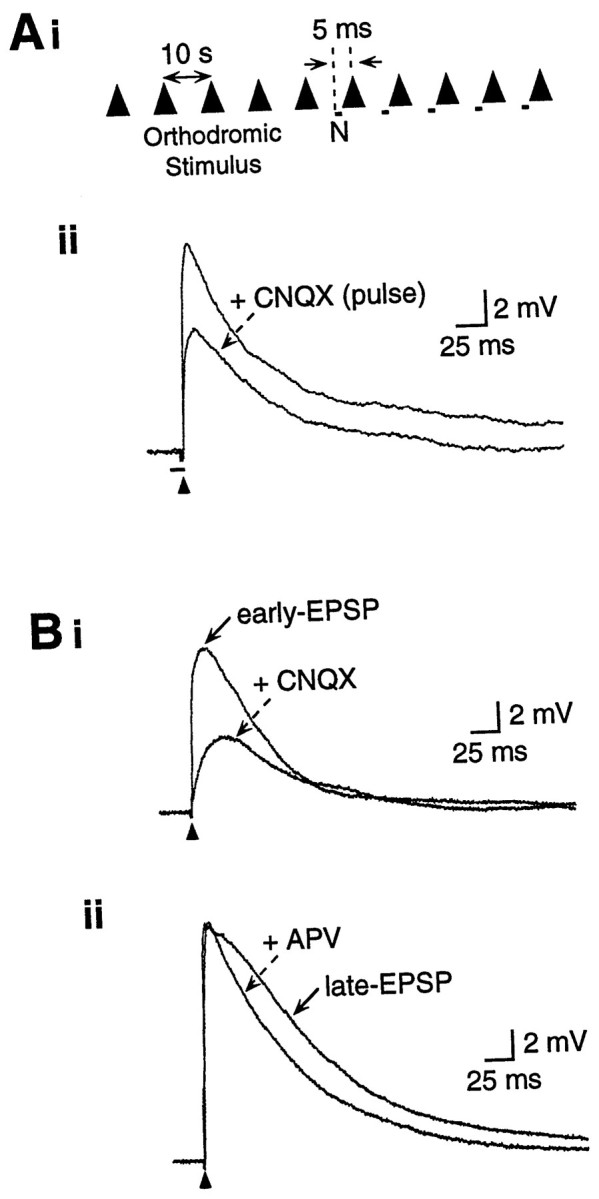
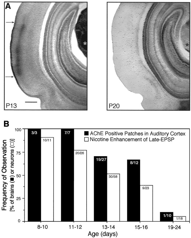
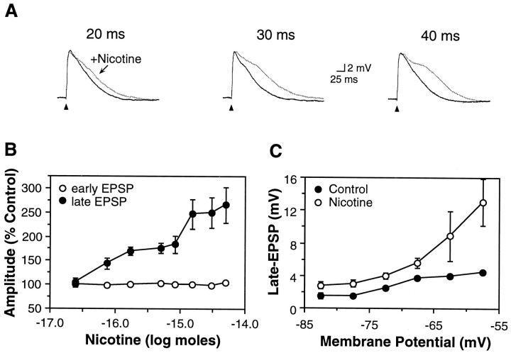
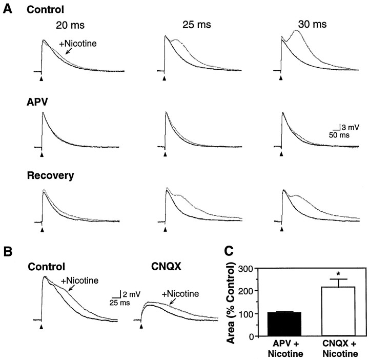
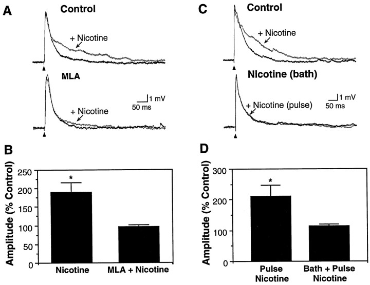
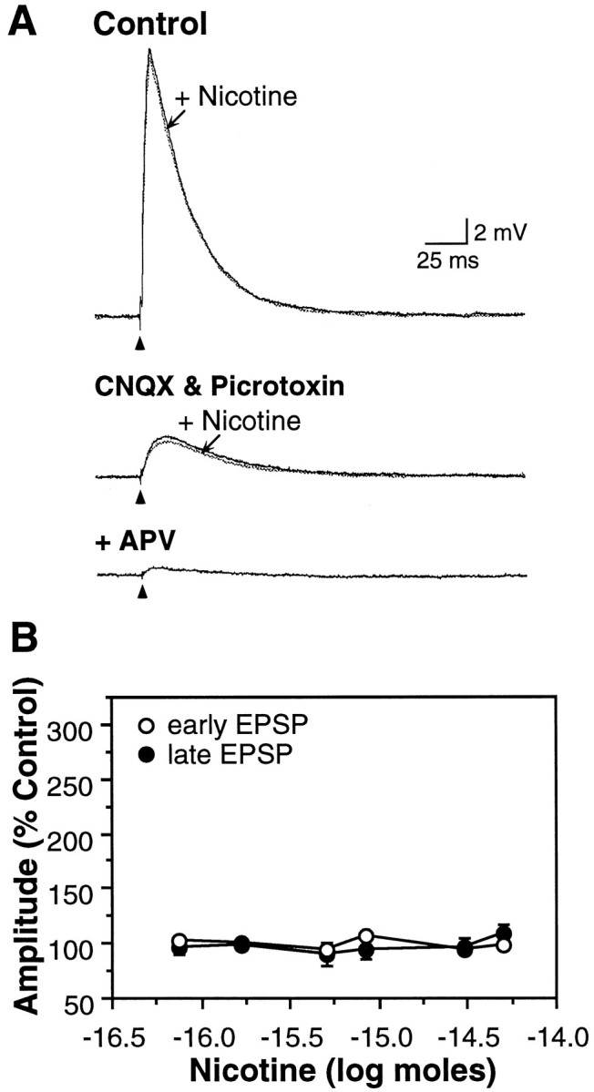
The neurotransmitters acetylcholine (ACh) and glutamate have been separately implicated in synaptic plasticity during development of sensory neocortex. Here we show that these neurotransmitters can, in fact, act synergistically via their actions at nicotinic ACh receptors (nAChRs) and NMDA receptors, respectively. To determine how activation of nAChRs modifies glutamatergic EPSPs, we made whole-cell recordings from visualized pyramidal neurons in slices of rat auditory cortex. Pulsed (pressure) ejection of nicotine onto apical dendrites selectively enhanced EPSPs mediated by NMDA receptors without affecting AMPA/kainate (AMPA/KA) receptor-mediated EPSPs. The enhancement occurred during a transient, postnatal period of heightened cholinergic function [neurons tested on postnatal day 8-16 (P8-16)], and not in the mature cortex (>P19). Three related findings indicated the mechanism of action: (1) The specific alpha7 nAChR antagonist methyllycaconitine citrate (MLA) blocked the effect of nicotine; (2) pulsed nicotine did not enhance postsynaptic depolarizations induced by iontophoretically applied NMDA; and (3) bath exposure to nicotine for several minutes produced apparent nAChR desensitization and precluded enhancement of EPSPs by pulsed nicotine. Together, the data suggest that nicotine acts at rapidly desensitizing, presynaptic alpha7 nAChRs to increase glutamate release onto postsynaptic NMDA receptors. The synergistic actions mediated by alpha7 nAChRs and NMDA receptors may contribute to experience-dependent synaptic plasticity in sensory neocortex during early postnatal life.


Figures

Fig. 1.
Fig. 1.
Experimental design and pharmacological profile of EPSPs. Ai, Synaptic potentials were elicited by a single stimulus pulse every 10 sec. After control responses, pressure-ejected nicotine (N) was paired for several trials, with each pressure pulse beginning 5 msec before the stimulus. Responses for each condition depicted in figures are averages of three to five traces. Aii, Orthodromic stimulus (arrowhead) elicited a glutamatergic EPSP in a layer III pyramidal neuron. Pressure ejection of CNQX (100 μm, 50 msec beginning 5 msec before the stimulus) reduced the peak EPSP amplitude, demonstrating that pressure-ejected drugs reach the synapse sufficiently quickly to affect the peak EPSP. Receptor antagonists are bath-applied in all other figures. Vm, −70 mV.B, Glutamatergic EPSPs are produced by activation of AMPA/KA and NMDA receptors. Bi, The peak EPSP (i.e., early EPSP) was significantly reduced by CNQX (20 μm in bath), indicating the involvement of AMPA/KA receptors. The late EPSP was subsequently reduced by APV (50 μm; data not shown). Vm, −66 mV. Bii, In another cell, the late/descending slope of the EPSP (i.e., late EPSP) was reduced by APV (50 μm), indicating the involvement of NMDA receptors. Note that APV did not reduce the peak EPSP amplitude. Vm, −70 mV.
Fig. 2.
Fig. 2.
Age-dependent expression of AChE patches and responsiveness of neurons to nicotine. A, Dense AChE staining (between arrows) occurred in layers I and III/IV of primary auditory cortex and in the auditory thalamus (*) of a P13 rat but disappeared by P20. Scale bar, 500 μm. B, The frequency of slices with AChE-positive patches declined with increasing age (correlation coefficient r = −0.91), as did the frequency of neurons whose synaptic activity was modified by nicotine (r = −0.98). Correlation coefficients were calculated using the product-moment method. Number of observations indicated within each column.
Fig. 3.
Fig. 3.
Nicotine selectively enhanced the late EPSP.A, Afferent stimulation elicited an EPSP (black traces) in a P16 pyramidal neuron. Pressure ejection of nicotine (25 μm, 20–40 msec pulse) produced a dose-dependent increase in the magnitude of the late EPSP without affecting the amplitude of the early EPSP (gray traces). Afferent stimuli delivered at 10 sec intervals; control traces were obtained between nicotine doses. Vm, −66 mV. B, Nicotine produced a dose-dependent increase in the late EPSP (•; n = 47; p < 0.05), but did not affect the amplitude of the early EPSP (○; p > 0.05). Average Vm, −67.8 ± 0.8 mV; neurons from P8–16 animals. C, Membrane depolarization increased the amplitude of the late EPSP (•, n = 14) and the degree to which nicotine enhanced the late EPSP (○).
Fig. 4.
Fig. 4.
Effect of nicotine on pharmacologically isolated EPSPs. A, Nicotine (25 μm, 20–30 msec pulse) produced a dose-dependent increase in late EPSP magnitude (Control; P13 pyramidal neuron). Pharmacological isolation of the AMPA/KA receptor-mediated early EPSP with APV (50 μm) revealed no effect of nicotine. Recovery followed wash-out of APV. Vm, −70 mV. B, The NMDA receptor-mediated late EPSP was isolated with CNQX (20 μm) and was enhanced by nicotine (25 μm, 30 msec). Recovery from CNQX was not attempted. Neuron from P16 rat; Vm, −64 mV. C, In nine cells with nicotine-induced enhancement of the late EPSP, the effects of nicotine were subsequently tested on pharmacologically isolated EPSPs (neurons from P8–16 animals). Nicotine had no effect on the isolated early EPSP (102.6 ± 7.3% of EPSP area in APV alone; p> 0.10; n = 5) but significantly increased the isolated late EPSP (to 214.5 ± 35.9% of EPSP area in CNQX alone;p < 0.01; n = 4).
Fig. 5.
Fig. 5.
The effect of nicotine was blocked by α7 nAChR antagonism and by persistent exposure to nicotine. A, Nicotine-induced (25 μm, 40 msec) enhancement of the late EPSP in a P8 neuron (top traces) was blocked by superfusion of the α7 receptor antagonist MLA (5 nm,bottom traces). Vm, −53 mV.B, In five neurons (age range, P8–13), nicotine enhancement of the late EPSP (to 188.1 ± 27.9% of control amplitude; p < 0.01) was prevented by MLA (5 nm; 95.5 ± 6.0% of control; p > 0.10). C, Enhancement of the late EPSP by pressure-pulse application of nicotine (25 μm, 30 msec; top traces) was prevented by bath application of a low concentration of nicotine (0.3 μm for 3 min,bottom traces). Vm, −68 mV.D, In five neurons (age range, P8–12), pulsed nicotine enhancement of the late EPSP (to 210.8 ± 37.1% of control;p < 0.05) was prevented by exposure to superfused nicotine (0.3–0.5 μm; 112.5 ± 8.5% of control;p > 0.10). Nicotine-induced enhancement was restored on washing for 6–8 min (220.5 ± 61.2%;n = 3).
Fig. 6.
Fig. 6.
Nicotine did not exert direct postsynaptic effects. A, Iontophoretic application of NMDA (15 nA, 3 sec; 50 mm) to a neuron from a P10 rat produced repeatable membrane depolarizations in low Ca2+/high Mg2+ ACSF. Concomitant pulse application of nicotine (25 μm; 20–100 msec) did not alter the magnitude of NMDA-induced responses. Vm, −70 mV.B, In six neurons (age range, P8–10), nicotine did not affect the amplitude of NMDA-induced membrane depolarizations, which averaged 11.7 ± 3.0 mV (peak depolarization) in the control condition and 10.7 ± 3.0 mV with a 50 msec pulse of nicotine (p > 0.10). C, Pulsed nicotine (marked by rectangle under baseline) in the absence of electrical afferent stimulation resulted in a small amplitude membrane depolarization. The nicotine-induced depolarization was prevented by superfusion of i, APV (50 μm, Vm, −61 mV); or ii, MLA (5 nm, Vm, −53 mV); but notiii, CNQX (20 μm, Vm, −61 mV). D, The nicotine-induced depolarization of Vm was voltage-sensitive and of greatest amplitude at depolarized potentials (n = 10 neurons; age P10–13).
Fig. 7.
Fig. 7.
Nicotine did not enhance EPSPs in the mature neocortex. A, Nicotine (10 μm, 50 msec) did not modify the EPSP in a P20 neuron. Superfusion of CNQX (20 μm) and picrotoxin (10 μm) isolated the late EPSP, which remained unaffected by nicotine (middle trace) and was blocked by APV (50 μm;lower trace). Increasing the stimulus intensity resulted in a larger (11 mV) isolated late EPSP in CNQX and picrotoxin that also was not affected by nicotine (data not shown). Vm, −77 mV. B, Nicotine did not affect EPSPs in P19–24 neurons (n = 18; p values > 0.10). Average Vm, −70.4 ± 1.5 mV.

References

    1. Agmon A, O’Dowd DK. NMDA receptor-mediated currents are prominent in the thalamocortical synaptic response before maturation of inhibition. J Neurophysiol. 1992;68:345–349. - PubMed
    1. Alkondon M, Rocha ES, Maelicke A, Albuquerque EX. Diversity of nicotinic acetylcholine receptors in rat brain. V. α-Bungarotoxin-sensitive nicotinic receptors in olfactory bulb neurons and presynaptic modulation of glutamate release. J Pharmacol Exp Ther. 1996;278:1460–1471. - PubMed
    1. Aramakis VB, Bandrowski AE, Ashe JH. Activation of muscarinic receptors modulates NMDA-receptor mediated responses in auditory cortex. Exp Brain Res. 1997;113:484–496. - PubMed
    1. Bear MF, Singer W. Modulation of visual cortical plasticity by acetylcholine and noradrenaline. Nature. 1986;320:172–176. - PubMed
    1. Bekkers J, Stevens C. NMDA and non-NMDA receptors are co-localized at individual excitatory synapses in cultured rat hippocampus. Nature. 1989;341:230–233. - PubMed

Publication types

MeSH terms

LinkOut - more resources