. 1998 Oct 15;18(20):8505-14.
doi: 10.1523/JNEUROSCI.18-20-08505.1998.

Mice lacking the beta3 subunit of the GABAA receptor have the epilepsy phenotype and many of the behavioral characteristics of Angelman syndrome

Affiliations

Mice lacking the beta3 subunit of the GABAA receptor have the epilepsy phenotype and many of the behavioral characteristics of Angelman syndrome

T M DeLorey et al. J Neurosci. .

Abstract

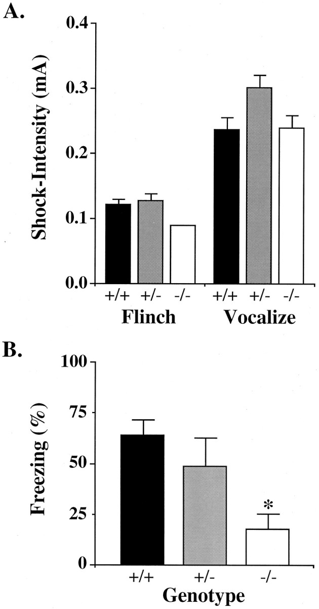
Angelman syndrome (AS) is a severe neurodevelopmental disorder resulting from a deletion/mutation in maternal chromosome 15q11-13. The genes in 15q11-13 contributing to the full array of the clinical phenotype are not fully identified. This study examines whether a loss or reduction in the GABAA receptor beta3 subunit (GABRB3) gene, contained within the AS deletion region, may contribute to the overall severity of AS. Disrupting the gabrb3 gene in mice produces electroencephalographic abnormalities, seizures, and behavior that parallel those seen in AS. The seizures that are observed in these mice showed a pharmacological response profile to antiepileptic medications similar to that observed in AS. Additionally, these mice exhibited learning and memory deficits, poor motor skills on a repetitive task, hyperactivity, and a disturbed rest-activity cycle, features all common to AS. The loss of the single gene, gabrb3, in these mice is sufficient to cause phenotypic traits that have marked similarities to the clinical features of AS, indicating that impaired expression of the GABRB3 gene in humans probably contributes to the overall phenotype of Angelman syndrome. At least one other gene, the E6-associated protein ubiquitin-protein ligase (UBE3A) gene, has been implicated in AS, so the relative contribution of the GABRB3 gene alone or in combination with other genes remains to be established.


Figures

Fig. 1.
Fig. 1.
Map of human chromosome 15 (top,H) and mouse chromosome 7 (bottom, M), indicating the arrangement of the UBE3A gene, the GABAA receptor gene cluster (GABRB3, GABRA5, GABRG3), and the P gene. The large deletion on maternal 15q11–13, indicated on the diagram with a dashed line, occurs in the majority of Angelman syndrome probands. The DNA region targeted for disruption in the gabrb3 gene knock-out mouse is indicated above the GABRB3 gene (Homanics et al., 1997). D15S541, SNRPN, and D15S144 are polymorphic (CA)nmicrosatellite markers used for determining the extent of the chromosomal deletion in humans. IC represents the region in which the “imprinting center” is found. Genes are presented in the diagram as the human homologs. The numbers on the mouse chromosome correlate to equivalent (syntenic) regions of human chromosomes.
Fig. 2.
Fig. 2.
Normal and abnormal background EEGs in both human and mouse. Comparisons of awake EEG between a normal human and individuals with different classes of Angelman syndrome.A, Normal, Segment of routine EEG on a normal 10-yr-old male with no seizures.AS-Deletion, Background EEG of a 9.5-yr-old male, large deletion AS case.AS-Non-Deletion, Background EEG of a 10-yr-old male with a UBE3A gene loss-of-function mutation. Both AS patients have been previously reported (Minassian et al., 1998) and fulfill consensus clinical criteria for AS (Williams et al., 1995). The deletion case was shown to have a large cytogenetically detectable deletion in chromosome 15q11–13. This was further confirmed using fluorescent in situ hybridization with probes D15S11 and GABRB3. A loss-of-function mutation was shown in the UBE3A case by Kishino et al. (1997). Routine EEG was performed using the international 10–20 electrode placement method on both AS patients and the age-matched normal child (for methods, see Minassian et al., 1998).Inset shows the location of the electrodes through which the tracings shown in this figure were obtained.F3-C3 indicates that the voltage of the C3 scalp electrode was subtracted from the F3 scalp electrode.C3-P3 indicates that the voltage from the P3 scalp electrode was subtracted from the C3 scalp electrode.B, Background EEGs from mouse littermates (gabrb3+/+ and gabrb3−/−) recorded simultaneously at 2 months of age. Electrodes were placed over right and left parietal cortex and referenced to an electrode placed in the nasal bone. Bottom two EEG traces are representative examples from a gabrb3−/− mouse before (b.) and after (a.) administration of ethosuximide (400 mg/kg). Ethosuximide effectively abolished interictal spiking and normalized EEG background.
Fig. 3.
Fig. 3.
Examples of gabrb3−/− and gabrb3+/− mouse EEG recordings during seizure episodes. A, Example of EEG-recorded ictal spikes in a 19-week-old gabrb3−/− mouse. These spikes, accompanied by strong head and forelimb myoclonic jerks, are seen frequently in gabrb3−/− mice and to a lesser extent in gabrb3+/− mice and not at all in gabrb3+/+, C57, or 129 mice.B, Example of EEG-recorded spike-wave discharges during facial and forelimb clonus in a 14 week old gabrb3−/− mouse.C, EEG recording of a gabrb3−/− (18-week-old) andD, gabrb3+/− (13-week-old) mouse taken during a generalized convulsive seizure in which both mice fell on their sides and exhibited vigorous forelimb and hindlimb clonus.
Fig. 4.
Fig. 4.
Performance of gabrb3 mouse genotypes and background mice strains in the step-through passive avoidance task. The three gabrb3 mouse genotypes along with the two progenitor strains (C57 and 129 mice) were trained and 48 hr later tested for retention of the learned task. Histogram of the mean time (seconds) to reenter dark chamber are presented for each mouse group, gabrb3+/+ (n = 18), gabrb3+/− (n = 27), gabrb3−/− (n = 12), C57 (n = 20), and 129 (n = 14). The gabrb3−/− mice were significantly different from gabrb3+/+ mice, p < 0.05. Error bars indicate the SEM. Asterisk identifies a significant difference from gabrb3+/+ mice, *p < 0.05.
Fig. 5.
Fig. 5.
Evaluation of pain perception and Pavlovian contextual fear conditioning in gabrb3 mouse genotypes.A, Pain perception assayed by behavioral response to a mild footshock. Histogram of mean milliamp current required to elicit the indicated behavioral response (“flinch” or “vocalization”) in the grouped mouse genotypes, gabrb3+/+ (n = 7), gabrb3+/− (n = 7), and gabrb3−/− (n = 6). The difference in behavioral response to shock between the three gabrb3 genotypes was not significant (p > 0.05). B, Pavlovian contextual fear conditioning assessed by ability to remember a mild footshock. Memory of a mild footshock was determined by measuring freezing time when the mouse was placed in a test cage in which 1 week previous it received a fear-conditioning mild footshock. Histogram of freezing scores are expressed as the mean of the percentage of total observations within genotype groups, gabrb3+/+ (n = 7), gabrb3+/− (n = 7), and gabrb3−/− (n = 6). The gabrb3−/− mice were significantly different from gabrb3+/+ mice, p < 0.05. Error bars indicate the SEM. Asterisk identifies significant difference from gabrb3+/+ mice, *p < 0.05.
Fig. 6.
Fig. 6.
Evaluation of motor activity levels in the gabrb3 mouse genotypes. A, Crossover activity was assessed by counting the number of times an individual mouse crossed the centerline of the test cage with all four paws. Data are presented as the mean (± SEM) within genotype groups, gabrb3+/+ (n = 7), gabrb3+/− (n = 7), gabrb3−/− (n = 6), of the number of crossovers made during an 8 min test period. The gabrb3−/− mice were significantly different from gabrb3+/+ mice, p = 0.01. B, Burst activity was determined by measuring the velocity of each mouse during a preshock and shock period. Baseline velocity was determined by dividing the distance the mouse traveled by the 20 sec period just before receiving a mild footshock. Shock velocity was determined by dividing the distance the mouse traveled by 2 sec (footshock duration). The gabrb3−/− mice were significantly more active than gabrb3+/+ mice. Error bars indicate the SEM. gabrb3+/+ (closed squares), gabrb3+/− (open triangles), gabrb3−/− (gray squares).Asterisks identify significant differences from gabrb3+/+, *p < 0.05, **p < 0.01.
Fig. 7.
Fig. 7.
Performance of gabrb3 mouse genotypes on a repeated motor coordination task. Mice were evaluated on the rotarod test once a day for 8 consecutive trial days. Data are presented as the mean (± SEM) of the time in which each mouse genotype was able to remain on a slowly rotating rod accelerated from 3.25 to 19 rpm over a 180 sec trial period, gabrb3+/+ (closed squares)n = 24, gabrb3+/− (open circles)n = 23, gabrb3−/− (open squares)n = 26. The gabrb3−/− mice exhibited significantly poorer performance on the rotarod task (trials 3–8) than that of the gabrb3+/+ mice (unpaired two-tailed t test). C57 (n = 24) and 129 (n = 24) were not significantly different in rotarod behavior to that of the gabrb3+/+ mice in trials 2–8 (data not shown). Error bars indicate the SEM. Asterisks identify significant differences from gabrb3+/+, *p < 0.05, **p < 0.01, ***p < 0.005.
Fig. 8.
Fig. 8.
Assessment of the rest–activity cycle of gabrb3 mouse genotypes by motion monitoring. Representative examples of recordings of rest–activity periods of the three different mouse genotypes (gabrb3+/+, gabrb3+/−, and gabrb3−/−) over a 2.5 d period. A 12 hr light/dark cycle was maintained during this period (bars at the bottom of the figure indicate the dark cycle). The activity units are arbitrary units based on integrated output voltage and plotted as a fraction of maximal output for each separate experiment.

References

    1. Bottani A, Robinson WP, DeLozier-Blanchet CD, Engel E, Morris MA, Schmitt B, Thun-Hohenstein L, Schinzel A. Angelman syndrome due to paternal uniparental disomy of chromosome 15: a milder phenotype? Am J Med Genet. 1994;51:35–40. - PubMed
    1. Boyd SG, Harden A, Patton MA. The EEG in early diagnosis of the Angelman (Happy Puppet) syndrome. Eur J Pediatr. 1988;147:508–513. - PubMed
    1. Buiting K, Saitoh S, Gross S, Dittrich B, Schwartz S, Nicholls RD, Horsthemke B. Inherited microdeletions in the Angelman and Prader-Willi syndromes define an imprinting centre on human chromosome 15. Nat Genet. 1995;9:395–400. - PubMed
    1. Chen C, Kim JJ, Thompson RF, Tonegawa S. Hippocampal lesions impair contextual fear conditioning in two strains of mice. Behav Neurosci. 1996;110:1177–1180. - PubMed
    1. Christian SL, Robinson WP, Huang B, Mutirangura A, Line MR, Nakao M, Surti U, Chakravarti A, Ledbetter DH. Molecular characterization of two proximal deletion breakpoint regions in both Prader-Willi and Angelman syndrome patients. Am J Hum Genet. 1995;57:40–48. - PMC - PubMed

Publication types

MeSH terms

Substances