. 1998 Oct 15;18(20):8539-49.
doi: 10.1523/JNEUROSCI.18-20-08539.1998.

Synaptic regulation of action potential timing in neostriatal cholinergic interneurons

Affiliations

Synaptic regulation of action potential timing in neostriatal cholinergic interneurons

B D Bennett et al. J Neurosci. .

Abstract

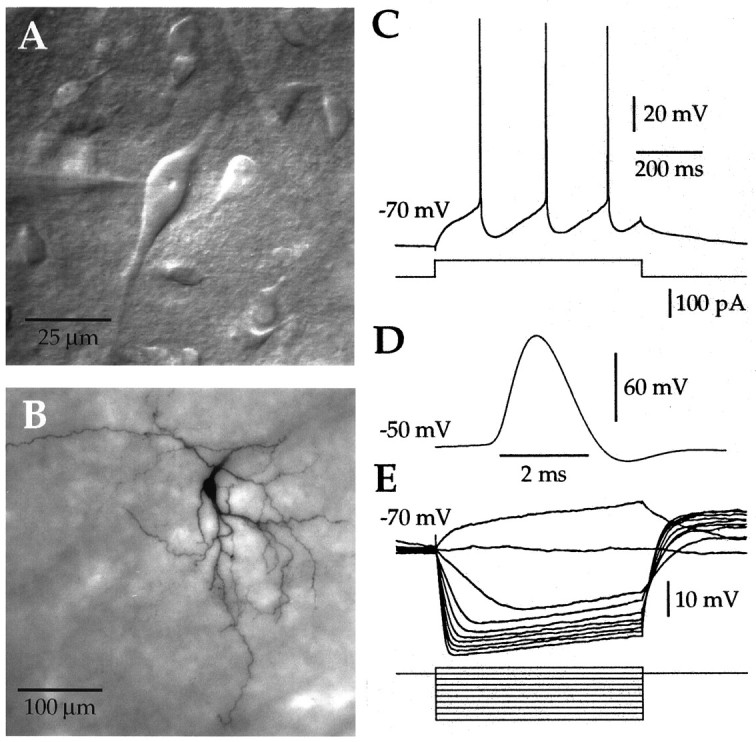
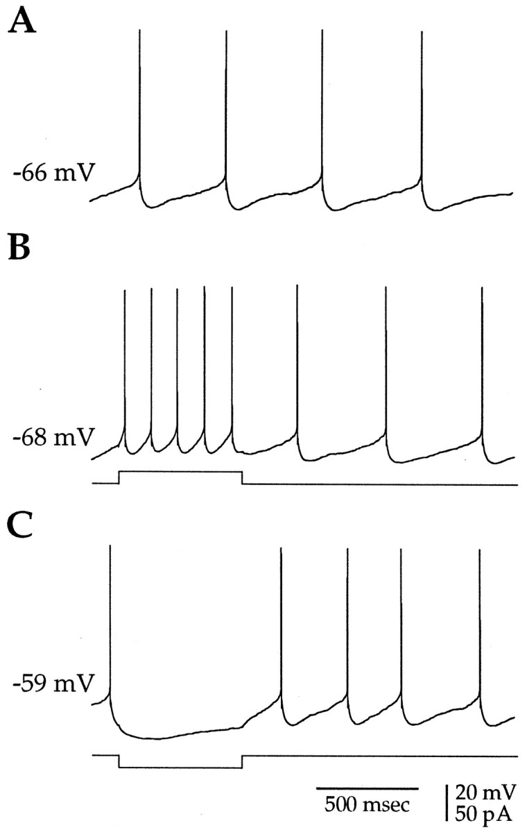
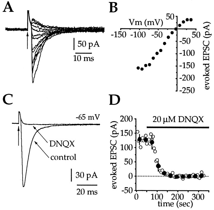
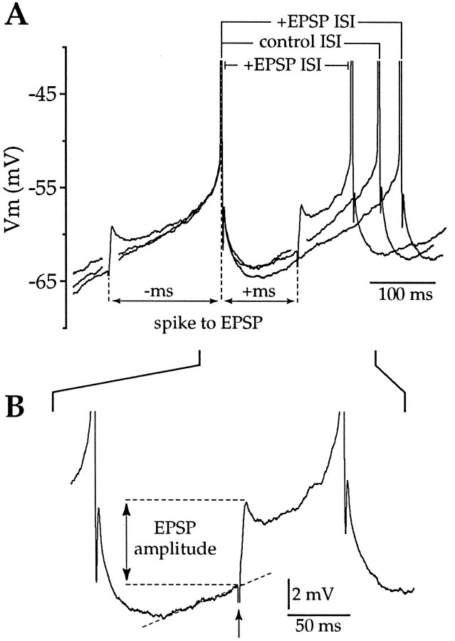
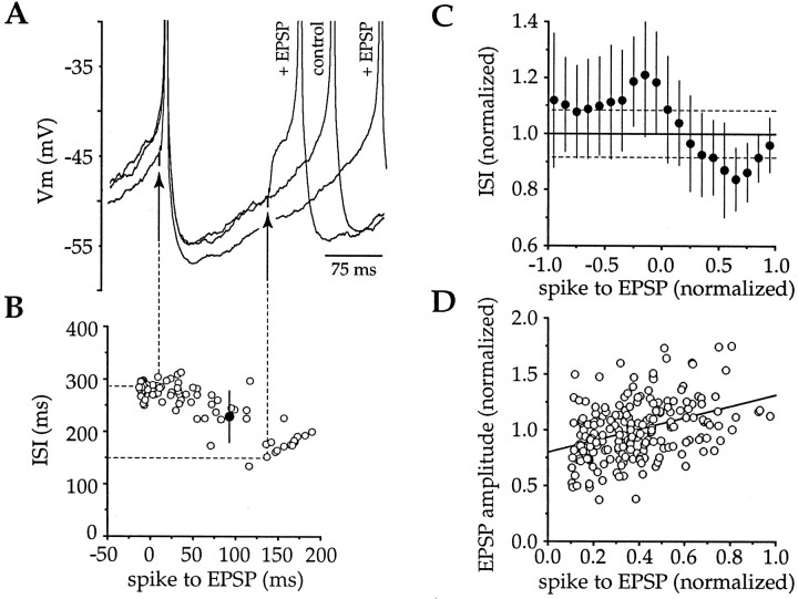
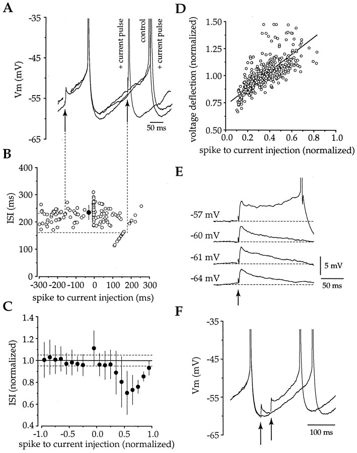
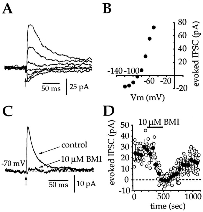
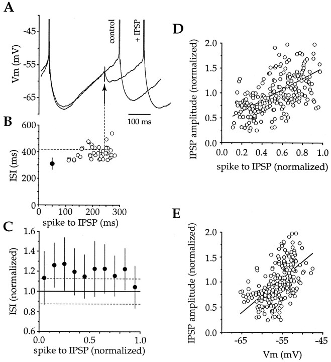
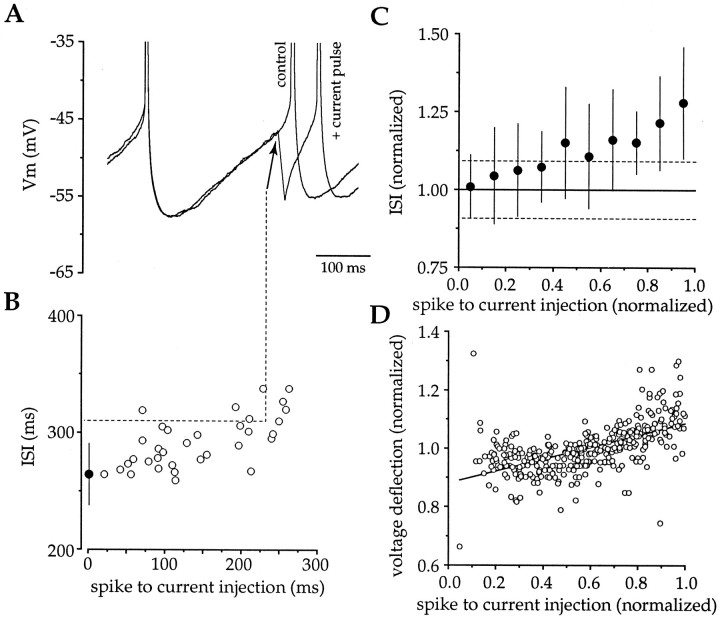
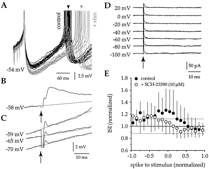
Action potentials in neostriatal cholinergic interneurons recorded in vivo are triggered by summation of two or three discrete synaptic depolarizations (Wilson et al., 1990). The ability and precision with which EPSPs and IPSPs regulate action potential timing was therefore investigated in vitro. Cholinergic interneurons were identified on the basis of morphological and electrophysiological characteristics in neostriatal slices taken from 2- to 3-week-old postnatal rats recorded at 24-26 degreesC. During periods of induced regular firing, intrastriatal stimuli were used to evoke pharmacologically isolated monosynaptic AMPA receptor-mediated EPSPs or GABAA receptor-mediated IPSPs. EPSPs evoked during the interspike interval (ISI) produced a phase-dependent decrease in the ISI, whereas IPSPs produced a phase-independent prolongation of the ISI. Injection of brief depolarizing currents mimicked the action of EPSPs and revealed an alteration in the input resistance during the ISI. In contrast to IPSPs, the ability of brief hyperpolarizing current injections to delay spike generation was phase-dependent. After blockade of GABAergic and glutamatergic synaptic transmission, stimuli failed to produce a detectable conductance change but could still prolong the subsequent ISI primarily through a D1 dopamine receptor-mediated enhancement of the afterhyperpolarization (AHP). Hence, EPSPs are ideally suited to provide a precise regulation of spike timing in cholinergic cells, whereas IPSPs are more likely to influence the overall level of excitability. The D1-mediated modulation of the AHP may contribute to the prolonged ISI seen in tonically active neurons in vivo in monkeys trained to respond to a sensory cue.


Figures

Fig. 1.
Fig. 1.
Morphological and physiological characterization of neostriatal cholinergic interneurons. A, An IR-DIC image of a neostriatal slice illustrating the characteristic appearance of giant interneurons. The large soma and thick primary dendrites are stereotypical features of cholinergic cells. B, Synthetic projection micrograph of a giant cell filled with biocytinin vitro and subsequently stained using standard techniques. In addition to the morphological features visible under IR-DIC optics, the secondary and higher order dendrites can be seen to branch, becoming fine-diameter structures. C, Depolarizing somatic current injection elicited regular spiking with each action potential, followed by a large-amplitude long-duration AHP.D, Action potentials were slow, with a width at half-amplitude always in excess of 1 msec. E, Injection of negative current caused an initial hyperpolarization, followed by a sag in the membrane potential. Subthreshold positive current injection produced a depolarizing ramp. These morphological and physiological features are characteristic of neostriatal cholinergic interneurons. InC–E, the initial membrane voltage is indicated to theleft of each trace. Time and current calibration for E are the same as inC.
Fig. 2.
Fig. 2.
Small-amplitude current injections induce regular spiking in spontaneously active cholinergic neurons. A, The majority (>80%) of cholinergic neurons were spontaneously activein vitro. B, Depolarizing somatic current injection (20 pA, 600 msec, 0.2 Hz) was used to elicit regular spiking with a stationary interspike interval. C, Hyperpolarizing current injection (−20 pA) caused a cessation in spontaneous firing, illustrating that the spiking in cholinergic neurons was readily controlled by small-amplitude current injections. The initial membrane potential is indicated to the leftof each trace.
Fig. 3.
Fig. 3.
Current–voltage and pharmacological characterization of excitatory synaptic inputs to cholinergic cells.A, A series of EPSCs evoked by intrastriatal stimulation after blockade of NMDA and GABAA receptors at holding potentials between −105 to +35 mV. Vertical arrows inA and C indicate the stimulus artifact.B, Current–voltage plot for the same cell inA revealed a reversal potential of approximately −5 mV. Each point is the mean peak amplitude of three EPSCs evoked at each of the holding potentials. C, At −65 mV, bath application of DNQX (20 μm) completely blocked the evoked inward current. Traces are averages of 25–30 individual trials. D, Time series for the effect of bath application of DNQX on evoked EPSC amplitude from the same cell inC (open circles, individual EPSCs;filled circles, mean ± SD of six sequential EPSCs). These data illustrate that intrastriatal stimulation in the presence of APV and BMI evokes a solely AMPA receptor-mediated inward current.
Fig. 4.
Fig. 4.
Measurements made to determine the phase dependence of the amplitude of postsynaptic potentials and voltage deflections produced by current injections and their effects on ISI. In this example, the effects of evoked EPSPs on spike timing are illustrated, but analogous measurements were made for all experiments in which the effect of EPSPs, IPSPs, and current injections on spike timing were investigated. A, The ISI was measured for all epochs in which there was no synaptic stimulus, and a mean control ISI was calculated for each neuron. The ISI was then measured for each epoch in which an EPSP was evoked, and the value was expressed as a proportion of the mean control ISI. The time from the first spike to the stimulus was measured and expressed as a proportion of the mean control ISI to give the phase. The phase had a negative value when the EPSPs were evoked before the first spike and a positive value when they fell after the first spike. B, An enlargement of the area indicated in A illustrates how the amplitudes of the EPSPs were measured. In an individual cell, the amplitude of all EPSPs evoked during the ISI were measured, the mean was calculated, and individual EPSPs were then expressed as a proportion of the mean. Note that in this example, the EPSP that occurs during the ISI does not trigger a spike immediately but causes a depolarization which persists, causing the cell to fire >50 msec after the stimulus, whereas the EPSP triggered before the first spike has a prolonged time course but decays back to baseline before the cell spikes.
Fig. 5.
Fig. 5.
Intrastriatal stimulation produces biphasic effects on spike timing. A, EPSPs were evoked at various times before and during the ISI to determine the effects of excitatory synaptic potentials on spike timing. EPSPs that were evoked before the first spike appeared to cause an increase in the subsequent ISI, whereas EPSPs evoked late in the ISI shortened the time between spikes by producing a depolarization from which action potentials were triggered. EPSPs could trigger spikes directly or by producing a depolarization that persisted, reaching threshold after the initial peak. B, Plot of the effects of all EPSPs from this cell (open circles, individual epochs; filled circle, mean ± SD of control ISI) revealed that stimuli before the first spike consistently increased the ISI, whereas stimuli during the first half of the ISI had progressively less effect, and stimuli evoked during the second half of the ISI were excitatory.C, Pooled data from seven neurons illustrate that the biphasic effect of intrastriatal stimulation was consistent across the population examined (filled symbols, mean ± SD for data binned at 0.1 ISI intervals). Stimuli produced a significant (dotted lines indicate 95% confidence interval) prolongation or reduction in the ISI, depending on when the stimulus was delivered. D, Plot of normalized time between the spike and the EPSP versus the normalized EPSP amplitude for all neurons (n = 7) revealed a significant correlation between these two parameters, demonstrating a phase-dependent increase in the EPSP amplitude during the ISI (slope, 0.516; r = 0.382; df = 206;p < 0.001).
Fig. 6.
Fig. 6.
Brief somatic current injections mimic the excitatory actions of EPSPs and reveal changes inRin during the ISI. A, Injection of brief large-amplitude positive current pulses (0.5 nA, 1 msec) produced membrane depolarizations of an equivalent amplitude to that produced by synaptic EPSPs. Depolarizations before the first spike in a pair did not alter the subsequent ISI, whereas positive current injections in the latter two-thirds of the ISI were excitatory and mimicked the actions of EPSPs. B, A plot of all epochs from the cell in A confirmed that depolarizing current injections were without effect when given before the first spike but shortened the time between spikes when applied in the latter two-thirds of the ISI. Open circles, Individual epochs;filled circle, mean ± SD of control ISI.C, Examination of pooled data (n = 12) revealed that these effects were consistent across the population, with only the excitatory effects present when depolarizing current injections were delivered during the latter two-thirds of the ISI (filled symbols, mean ± SD for data binned at 0.1 ISI; dotted lines are 95% confidence intervals).D, Plot of phase of the current injection versus the normalized voltage deflection revealed large changes in the apparent input resistance of the neuron during the ISI (slope, 0.790;r = 0.729; df = 451; p < 0.001). E, F, Both EPSPs (E) and voltage deflections produced by current injections (F) elicit voltage-dependent prolonged depolarizations, indicative of the recruitment of a subthreshold regenerative inward current.
Fig. 7.
Fig. 7.
Current–voltage and pharmacological characterization of inhibitory synaptic inputs to cholinergic cells.A, After blockade of AMPA and NMDA receptors, intrastriatal stimulation at holding potentials between −120 and −60 mV evoked a voltage-dependent IPSC. Each trace is an individual IPSC. B, Current–voltage relationship for the cell in A. Each point represents the mean of three IPSCs evoked at each holding potential and indicates a reversal potential of approximately −88 mV, which is consistent with the value predicted by the Nernst equation. C, Application of 10 μm bicuculline completely blocked the evoked IPSC.Traces are averages of 30–50 individual IPSCs.D, Time series from the same cell in Cillustrates a reversible blockade of the evoked IPSCs after application and washout of bicuculline (open circles, individual IPSCs; filled circles, mean ± SD of six sequential IPSCs).
Fig. 8.
Fig. 8.
IPSPs produce a phase-independent prolongation of the ISI. A, Intrastriatal stimulation after blockade of AMPA and NMDA receptors elicited a GABAA receptor-mediated IPSP that delayed the subsequent spike. B, The ability of the IPSP to increase the ISI was independent of when the IPSP occurred during the ISI (open circles, individual epochs; filled circle, mean ± SD of control ISI).C, Examination of five neurons revealed that synaptically evoked IPSPs produced a prolongation of the ISI, irrespective of when the IPSP occurred during the ISI (filled circles, mean ± SD binned at 0.1 ISI; dotted lines indicate 95% confidence limits).D, E, Plots of normalized time from the spike to the IPSP versus the normalized IPSP amplitude (slope, 1.096;r = 0.577; df = 235; p < 0.001) or membrane potential at which the IPSP was evoked (slope, 0.067; r = 0.551; df = 235;p < 0.001). These data revealed the amplitude of the IPSP, as detected at the soma, was strongly dependent on when the IPSP occurred during the ISI.
Fig. 9.
Fig. 9.
Brief somatic hyperpolarizations delay spiking and confirm that Rin changes during the ISI.A, Injection of large-amplitude brief-duration negative current (−1 nA, 1 msec) produced a membrane hyperpolarization and delayed action potential generation. B, Examination of all epochs from the same cell in A demonstrated that the efficacy of somatic hyperpolarizations in delaying spiking were dependent on when the pulse was delivered during the ISI (open circles, individual epochs; filled circle, mean ± SD of control ISI). C, Pooled data (n = 8) illustrate that the efficacy of somatic hyperpolarization in delaying spike generation was observed across the population (filled circles, mean ± SD binned at 0.1 ISI; dotted lines indicate 95% confidence intervals). D, Plot of data from all cells for the relationship between phase and normalized voltage deflection illustrates the change in Rin taking place during the ISI (slope, 0.205; r = 0.582; df = 347; p < 0.001).
Fig. 10.
Fig. 10.
Prolongation of the ISI is attributable to D1 dopamine receptor-mediated enhancement of the AHP. A, A family of traces from a single neuron in which stimuli were delivered before the first spike in the presence of DNQX, APV, and BMI (gray) or no stimulus was given (black). Arrow indicates the mean ISI in each case. The stimulus causes a prolongation of the ISI by increasing the amplitude and duration of the AHP. B, An enlarged trace illustrates an EPSP evoked in the same neuron as in A, before application of DNQX.C, After blockade of the evoked AMPA receptor-mediated EPSP with DNQX (20 μm) and in the presence of BMI (10 μm) and APV (50 μm), the same stimulus fails to evoke any detectable synaptic response. D, Stimuli applied over a large range of membrane potentials in voltage-clamp recordings confirm that in the presence of DNQX, APV, and BMI no detectable conductance change is induced by intrastriatal stimulation. Recording in D made with a cesium and QX-314-containing electrode solution. E, Intrastriatal stimuli presented before or soon after a spike caused a prolongation of the ISI in the presence of APV, BMI, and DNQX (n = 8) (filled symbols). The prolongation was blocked by application of 10 μm SCH-23390 (n= 7) (open symbols), a D1 receptor antagonist (symbols, mean ± SD for data binned at 0.1 ISI intervals; dotted lines indicate 95% confidence limits).

References

    1. Adelman WJ, Jr, French RJ. Blocking of the squid axon potassium channel by external caesium. J Physiol (Lond) 1978;276:13–25. - PMC - PubMed
    1. Agard DA, Hiraoka Y, Shaw P, Sedat JW. Fluorescence microscopy in three dimensions. Methods Cell Biol. 1989;30:353–377. - PubMed
    1. Aosaki T, Kawaguchi Y. Actions of substance P on rat neostriatal neurons in vitro. J Neurosci. 1996;16:5141–5153. - PMC - PubMed
    1. Aosaki T, Graybiel AM, Kimura M. Effect of the nigrostriatal dopamine system on acquired neural responses in the striatum of behaving monkeys. Science. 1994a;265:412–415. - PubMed
    1. Aosaki T, Tsubokawa H, Ishida A, Watanabe K, Graybiel AM, Kimura M. Responses of tonically active neurons in the primate’s striatum undergo systematic changes during behavioral sensorimotor conditioning. J Neurosci. 1994b;14:3969–3984. - PMC - PubMed

Publication types

MeSH terms

LinkOut - more resources